 Log In
Search




 Open Access
 yyh.8@163.com
Received April 16, 2026; Accepted June 1, 2026; Published June 12, 2026
Open Accessyyh.8@163.comReceived April 16, 2026; Accepted June 1, 2026; Published June 12, 2026
Neuraxial anesthesia is often challenging in patients with obesity, spinal deformity, prior spinal surgery, ankylosing spondylitis, or of advanced age, as conventional landmark-based techniques frequently fail in this population. Ultrasound guidance has improved puncture accuracy; however, commonly used approaches may still be limited by patient positioning and the side from which the operator prefers to work. We report five cases of extremely difficult neuraxial anesthesia in which the procedure was successfully performed using an ultrasound-guided paramedian sagittal long-axis out-of-plane approach. This technique allows continuous visualization of the lamina and the anterior–posterior complex, providing a short and direct puncture path with real-time guidance. All procedures were completed successfully on the first attempt, with short puncture times and no anesthesia-related complications. These cases suggest that the ultrasound-guided long-axis out-of-plane approach is a safe, effective, and flexible option for managing difficult neuraxial anesthesia.
Spinal anesthesia is commonly used for lower extremity, pelvic and lower abdominal surgery. Traditional blind probing relies on well-defined anatomical landmarks; however, in patients with obesity, spinal deformity, history of spinal surgery, ankylosing spondylitis, or degenerative spinal changes, optimal patient positioning for the procedure is often difficult to achieve. This can lead to puncture failure or an increased risk of complications [1-3]. Ultrasound visualization provides real-time image guidance, improving accuracy and safety [4-6]. In ultrasound-guided spinal puncture, the short-axis plane and the paramedian sagittal oblique plane (a long-axis plane) are two common approaches [7-9]. The latter can clearly visualize multiple segments of the lamina, ligamentum flavum, and dural sac (i.e., the “anterior-posterior complex”) simultaneously [10]. However, it is more positionally demanding, as right-handed operators typically require the patient to lie on their left side. The long-axis in-plane technique allows direct visualization of the needle trajectory but is often limited by patient positioning and operator handedness (Figure 1A). In contrast, the long-axis out-of-plane technique is free from these constraints, allows real-time needle tip tracking, and is ideally suited for complex cases with unclear landmarks, narrow interspaces, or difficult positioning (Figure 1B). This paper reports five successful cases of difficult neuraxial anesthesia using the ultrasound-guided long-axis out-of-plane approach.

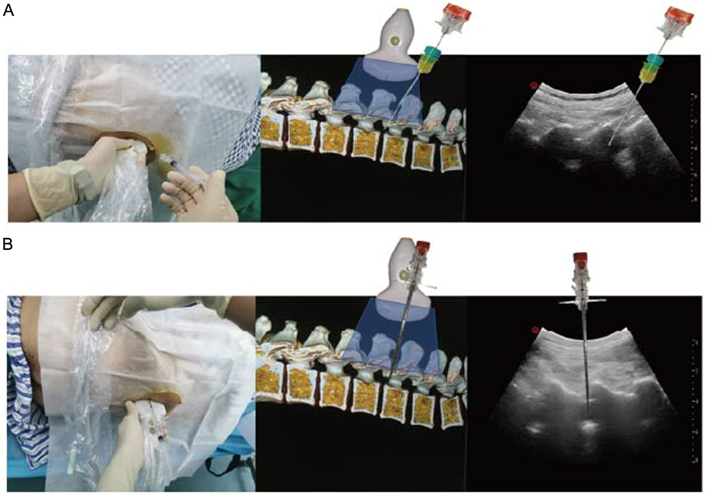
Figure 1. Comparison of long-axis in-plane and out-of-plane ultrasound-guided techniques. Compared with short-axis scanning, the longaxis view provides clearer visualization of intraspinal structures and allows continuous display of multiple interlaminar spaces, facilitating identification of the widest or most distinct acoustic window. The long-axis in-plane technique (A) enables direct visualization of the needle trajectory but is limited by patient positioning and operator handedness. In contrast, the long-axis out-of-plane technique (B) is not constrained by these factors, allows tracking of needle tip advancement, and is particularly suitable for patients with restricted positioning or narrow intervertebral spaces.
2.1 Case 1
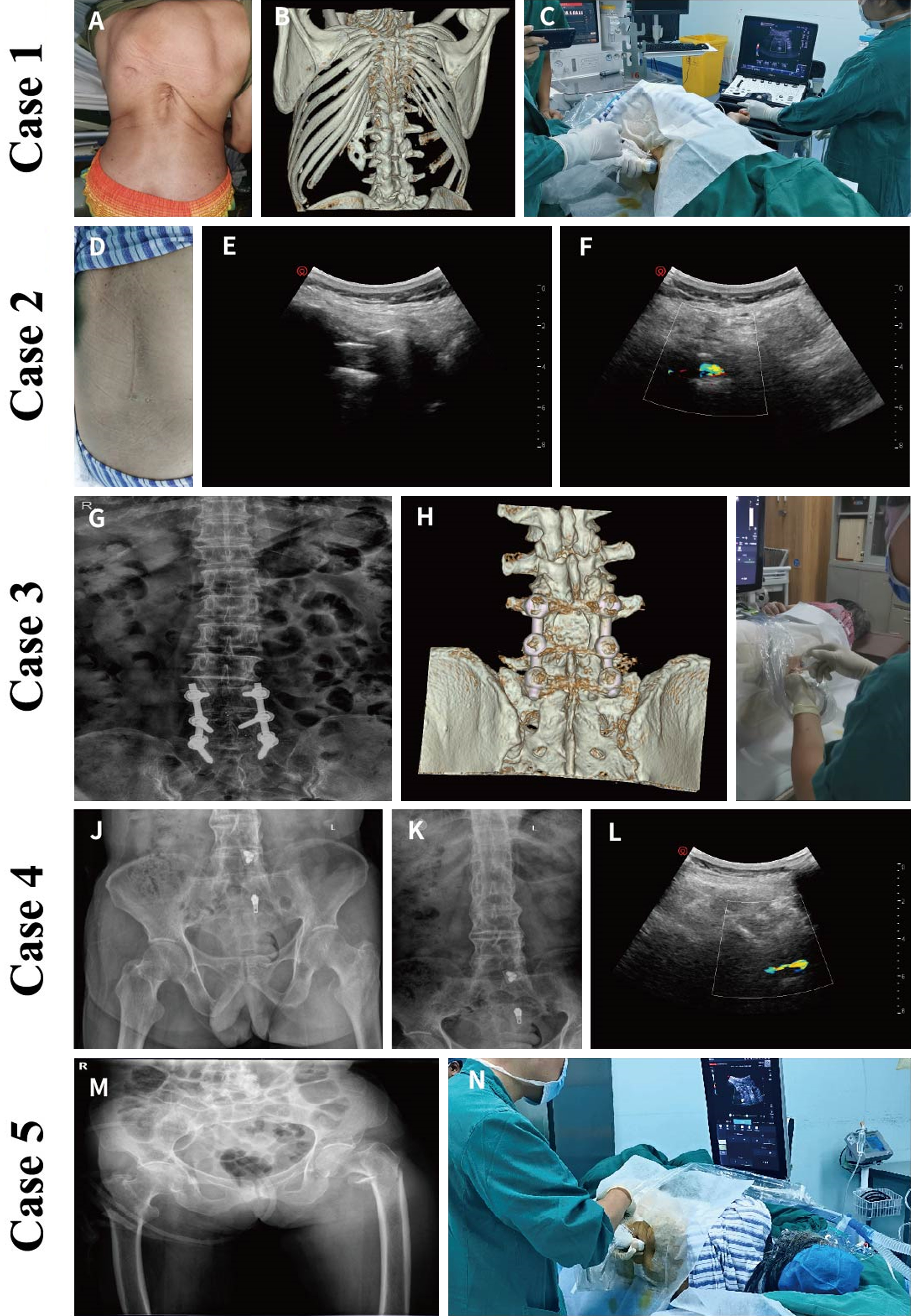
Figure 2. Ultrasound-guided long-axis out-of-plane neuraxial procedures in patients with challenging spinal conditions (Cases 1-5). (A-C) Case 1: A patient with chronic obstructive pulmonary disease and cor pulmonale underwent transurethral cystolithotripsy (A). Threedimensional reconstruction revealed a congenital thoracic deformity (B). Feasible puncture windows were identified at L4–L5 and L5–S1. With the patient in the left lateral position, combined spinal–epidural anesthesia was successfully performed at L4–L5 using an ultrasound-guided long-axis out-of-plane approach (C). (D-F) Case 2: A parturient with a history of lumbar internal fixation underwent cesarean section (D). Ultrasound scanning demonstrated that the anterior–posterior complex was widest at the L2–L3 interspace (E). Doppler imaging in the short-axis view showed blood flow signals during injectate administration (F). (G-I) Case 3: A patient with a history of L4–S1 lumbar internal fixation presented with left-sided lumbosacral radicular pain (G). Three-dimensional reconstruction demonstrated the internal fixation hardware (H). A puncture window was identified at L3–L4. With the patient in the right lateral position, a diagnostic lateral recess injection was performed at the L3–L4 interspace using an ultrasound-guided long-axis out-of-plane approach (I). (J-L) Case 4: A patient with a left intertrochanteric femoral fracture underwent proximal femoral nail antirotation surgery (J) and had concomitant ankylosing spondylitis (K). With the patient in the right lateral position, spinal anesthesia was performed at the L2–L3 interspace using an ultrasound-guided long-axis out-of-plane approach. Doppler signals generated by injectate spread were observed on the long-axis view (L). (M, N) Case 5: A patient with a left intertrochanteric femoral fracture underwent proximal femoral nail antirotation surgery (M). With the patient in the right lateral position, combined spinal–epidural anesthesia was successfully performed at the L5–S1 interspace using an ultrasound-guided long-axis out-of-plane approach (N).
2.2 Case 2
A 34-year-old obese parturient (160 cm, 85 kg, BMI 33.2 kg/m2) at 39 weeks’ gestation had a history of lumbar internal fixation and schizophrenia. Previous cesarean section had required general anesthesia. Using laminar long-axis imaging combined with transverse process localization, ultrasound scanning identified the widest anterior–posterior complex at L2–L3. Spinal anesthesia was successfully performed via a long-axis out-of-plane approach within 4 minutes, achieving a T6 sensory level. Doppler ultrasound confirmed intrathecal injection (Figure 2D-F).
2.3 Case 3
A 69-year-old patient (158 cm, 57 kg, BMI 22.8 kg/m2) with prior L4–S1 internal fixation presented with refractory left lumbar radicular pain. Under ultrasound guidance, an epidural puncture was performed at L3–L4 using the long-axis out-of-plane technique, followed by injection of 5 mL of anti-inflammatory and analgesic medication. The patient experienced immediate and significant pain relief (Figure 2G-I).
2.4 Case 4
A 64-year-old male (170 cm, 55 kg, BMI 19.0 kg/m2) with ankylosing spondylitis and a left intertrochanteric femoral fracture underwent a fascia iliaca compartment block followed by spinal anesthesia. Using ultrasound guidance in the right lateral position, spinal anesthesia was successfully achieved at L2–L3 via a long-axis out-of-plane approach. Doppler signals confirmed injectate spread, and the sensory block reached T8 without complications (Figure 2J-L).
2.5 Case 5
A 96-year-old patient (150 cm, 40 kg, BMI 17.8 kg/m2) with chronic lung disease and a left intertrochanteric fracture posed significant anesthetic challenges. Ultrasound scanning showed that only the L5–S1 anterior–posterior complex was clearly visible. Combined spinal–epidural anesthesia was successfully performed at this level using a long-axis out-of-plane approach. The puncture took 5 minutes, achieved a T10 block, and was well tolerated (Figure 2M, 2N; see Supplementary Video 1).
This case series includes patients with multiple factors associated with difficult spinal anesthesia, such as spinal deformity, obesity, prior spinal fixation surgery, ankylosing spondylitis, and advanced age. In these populations, conventional landmark-based puncture is often limited by obscure anatomical landmarks, inaccurate localization, and repeated needle attempts [1, 3]. Ultrasound-guided techniques—particularly the long-axis out-of-plane approach—offer an effective solution by enabling direct visualization of the interlaminar space, ligamentum flavum, and dural sac (see Supplementary Video 2) [8, 10]. This approach is not constrained by operator handedness or patient positioning and provides a short, direct puncture path.
The ultrasound-guided long-axis out-of-plane technique has several notable advantages. First, it allows superior visual localization. Compared with short-axis imaging, the long-axis view clearly delineates the spinal canal and enables continuous visualization of multiple interlaminar spaces, facilitating identification of the widest or most distinct acoustic window, especially in patients with absent or distorted surface landmarks [8, 10]. Second, it provides real-time guidance and confirmation. Needle advancement can be dynamically observed as it passes through the ligamentum flavum and dura mater. Correct needle tip placement can be further confirmed by anterior displacement of the dural sac or color Doppler signals generated by saline or drug injection, improving puncture accuracy and safety [9]. Third, this technique aids accurate interspace identification. Laminar oblique scanning combined with transverse process localization can reliably identify the target intervertebral level, which is particularly important in patients with altered spinal anatomy [9, 10]. Finally, in this small series of five patients, the technique was completed successfully on the first attempt in all cases, with short puncture times and no anesthesia-related complications. These preliminary findings suggest the procedure may be feasible and well-tolerated, although further studies with larger sample sizes are needed to evaluate its success rate and safety more definitively.
Several technical considerations are essential for successful application. A low-frequency convex probe is typically used, with paramedian sagittal oblique scanning to align the ultrasound beam parallel to the lamina and obtain an optimal view of the anterior–posterior complex. An out-of-plane needle insertion technique is employed, with needle depth tracked by subtle probe adjustments or the needle trajectory method. In difficult situations, such as when only a single interlaminar space is visible or when severe osteophyte formation is present, probe angulation may need adjustment, and epidural saline injection can assist in confirming loss of resistance and dural sac movement. However, ultrasound penetration is limited in cases of heavy calcification or extensive bony obstruction, underscoring the need for adequate operator training and experience.
In conclusion, this case series of five patients with complex spinal conditions demonstrates that the ultrasound-guided long-axis out-of-plane approach was successfully performed on the first attempt in all cases, with short puncture times and no anesthesia-related complications. These preliminary findings suggest the technique may be a feasible alternative for difficult neuraxial anesthesia, although its efficacy and safety compared with traditional methods cannot be determined from this small series. Further large-scale, controlled studies are warranted to validate these observations and define its potential role in clinical practice.
Author contributions
Liangqing Lin and Qinghua Wu conceptualized the study and designed the methodology. Pinhui Ke and Chunlan Lin were responsible for the data curation and formal analysis. Yaohua Yu performed the investigation and validated the results. All authors contributed to writing the original draft, reviewing, and editing the manuscript. All authors have read and agreed to the published version of the manuscript.
Funding
This research was funded by Putian Science and Technology Program 2025 (2025NJYL026).
Data availability
The individual deidentified participant data can be accessed from the corresponding author (Yaohua Yu, yyh.8@163.com) upon reasonable request.
Ethics approval and consent to participate
The study was approved by the Ethics Committee of The First Hospital of Putian (Approval No. 2026-026), and written informed consent was obtained from all participants or their legal representatives.
Consent for publication
All the authors have read and approved the final version of the manuscript and have consented to its submission for publication. The manuscript is not under consideration for publication elsewhere.
Competing interests
The authors declare that they have no competing interests.
Acknowledgements
Not applicable.
[2] Sivakumar RK, Karmakar MK. Spinal sonography and central neuraxial blocks. Best Pract Res Clin Anaesthesiol. 2023 Jun;37(2):209-242. https://doi.org/10.1016/j.bpa.2023.04.008
[3] Kamimura Y, Yamamoto N, Shiroshita A, Miura T, Tsuji T, Someko H, et al. Comparative efficacy of ultrasound guidance or conventional anatomical landmarks for neuraxial puncture in adult patients: A systematic review and network meta-analysis. Br J Anaesth. 2024 May;132(5):1097-1111. https://doi.org/10.1016/j.bja.2023.09.006
[4] Ke X, Wu Y, Yang R, Zhou Y, Zhang M, Liu H, et al. Real-time ultrasound-guided intrathecal delivery of nusinersen in adult patients with spinal muscular atrophy and complex spinal anatomy. Pain Physician. 2025 Nov;28(6):E695-E703.
[5] Paul A, Borkar A. Ultrasound-guided neuraxial anaesthesia for vaginal hysterectomy in a patient with rheumatoid arthritis. Cureus. 2023 Feb 16;15(2):e35064. https://doi.org/10.7759/cureus.35064
[6] Creaney M, Mullane D, Casby C, Tan T. Ultrasound to identify the lumbar space in women with impalpable bony landmarks presenting for elective caesarean delivery under spinal anaesthesia: A randomised trial. Int J Obstet Anesth. 2016 Dec;28:12-16. https://doi.org/10.1016/j.ijoa.2016.07.007
[7] Sivakumar RK, Areeruk P, Mok LYH, Samy W, Karmakar MK. Real-time ultrasound-guided spinal injection using a transverse in-plane dependent technique: A case series. A A Pract. 2022 Jul 11;16(7):e01596. https://doi.org/10.1213/XAA.0000000000001596
[8] Chin KJ, Chan VWS, Ramlogan R, Perlas A. Real-time ultrasound-guided spinal anesthesia in patients with a challenging spinal anatomy: Two case reports. Acta Anaesthesiol Scand. 2010 Feb;54(2):252-255. https://doi.org/10.1111/j.1399-6576.2009.02112.x
[9] Karmakar MK, Li X, Ho AMH, Kwok WH, Chui PT. Real-time ultrasound-guided paramedian epidural access: Evaluation of a novel in-plane technique. Br J Anaesth. 2009 Jun;102(6):845-854. https://doi.org/10.1093/bja/aep079
[10] Grau T, Leipold RW, Horter J, Conradi R, Martin EO, Motsch J. Paramedian access to the epidural space: The optimum window for ultrasound imaging. J Clin Anesth. 2001 May;13(3):213-217. https://doi.org/10.1016/S0952-8180(01)00245-8
ISSN: 2957-5443
Volume 4, Issue 2
June 2026
Pages: 149-224


 Log In

 On This Page
 CITE
CITE