 Log In
Search




 Open Access
 songzhijing2020@163.com
Zhijing Song, School of Clinical Chinese Medicine, Gansu University of Chinese Medicine, No. 35 Dingxi East Road, Chengguan District, Lanzhou 730000, Gansu, China. E-mail: songzhijing2020@163.com.
Open Accesssongzhijing2020@163.comZhijing Song, School of Clinical Chinese Medicine, Gansu University of Chinese Medicine, No. 35 Dingxi East Road, Chengguan District, Lanzhou 730000, Gansu, China. E-mail: songzhijing2020@163.com.
Sepsis is a systemic inflammatory response syndrome triggered by infection. It presents with simultaneous overactive immune activation and immune paralysis, leading to a cytokine storm, tissue injury, and multiple organ failure. The molecular mechanisms and pathological characteristics of the septic cytokine storm are comprehensively interpreted from multiple perspectives, including immune regulation, signaling pathways, cellular interactions, metabolic state reprogramming, and microcirculatory dysfunction. The cross-activation of signaling pathways, including NF-κB, JAK/STAT, mitogen-activated protein kinase (MAPK), and NLRP3 inflammasome, underlies the core mechanism for the enhancement of the inflammatory response via continued release of proinflammatory cytokines and also is involved in maintaining immune balance. In the latestage immunosuppressive phase, Treg cells, myeloid-derived suppressor cells (MDSCs) and regulatory macrophages play pivotal roles in reestablishing host homeostasis through negative feedback, and their dysfunction is closely associated with secondary infections and high mortality. This review consolidates the latest developments in precision medicine and multidimensional biomarkers, emphasizing the prospective value of several markers such as lactate and CD64, pentraxin 3 (PTX3), and BMP9 for early diagnosis and rognosis evaluation. Moreover, it reviews novel immunomodulators, for instance, bee venom peptides, GDF15, N-(1,3,4-oxadiazol-2-yl) guanidine (NWG), 4-octyl itaconate (4-OI), and nanoparticle drug delivery systems that inhibit inflammatory signaling cascades, restore energy metabolism, and provide organ protection. Taken together, the pathogenic mechanisms of sepsis are non-linear and dynamic, which may render single-target therapy insufficient for successful management. Personalized interventions targeting multi-target network regulation based on omics and AI models may accomplish precision monitoring and stratified treatment of inflammatory responses, also providing new theoretical and clinical strategies to reduce sepsis-related mortality.
Sepsis is an infection-induced systemic inflammatory response syndrome. Overactivation of the immune system during its progression results not only in hyper-inflammatory responses but may also be associated with immunosuppression, known as immune paralysis, which makes treatment difficult. However, therapies to decrease immune activation may have deleterious effects on patients who are immunoparalyzed due to viral infection or sepsis [1]. Clinically, sepsis still has a high incidence and mortality, and remains a major public health problem worldwide. In 2017, the global number of new sepsis cases was estimated at 48.9 million (95% UI: 38.9–62.9), with an estimated 11 million sepsis-related deaths (95% UI: 10.1–12.0) [2]. The challenging development of existing therapies and their limited benefits underscore the pressing need for early diagnosis and targeted treatments. Among these, the cytokine storm is the most important characteristic of sepsis. It refers to an overwhelming shift toward pro-inflammatory responses over anti-inflammatory responses, resulting in systemic inflammatory amplification and immune homeostasis. High levels of serum ferritin were closely associated with increased formation of neutrophil extracellular traps (NETs) and aggravation of lung injury in sepsis patients, suggesting that ferritin might become a significant marker for assessing inflammation severity and immune imbalance. There is evidence that its level is closely associated with overall prognosis in sepsis patients [3].
Although therapeutic strategies for sepsis have greatly improved and may include antimicrobial therapy, fluid resuscitation, and supportive organ therapy in clinical practice, single-target intervention is not compatible with the complex super-network exhibited by the cytokine storm. The intercellular signaling pathways in sepsis represent a coordinated immunometabolic cross-talk. Studies have demonstrated that hepatocyte mitochondria are metabolically linked to TREM2-expressing liver macrophages, and disruption of this coordination may be further amplified during inflammatory responses and organ damage. This indicates that intercellular metabolic communication may represent a novel point of entry for precision interventions [4]. Additionally, GABA regulation, metabolic reprogramming, and the inflammatory macrophage response mediated by protein succinylation are also implicated, which complicates the non-linear dynamics of the pathophysiological mechanisms in sepsis. This complexity explains why the therapies developed so far have such narrow application ranges. Furthermore, the crosstalk within the inflammatory network, negative feedback regulation, and immune suppression in late-phase sepsis induce a transition from excessive inflammation to an immunodeficiency state, which predisposes patients to secondary infections and death. This also implies that early in the course of treatment, blood cultures probably have poor sensitivity for diagnosing patients with severe sepsis, especially if empirical antimicrobial therapy has been initiated [5].
In light of the above background, a systematic study of the multi-center network mechanisms in the sepsis cytokine storm, as well as immune regulation features and possible intervention strategies, has important theoretical and clinical implications. This review summarizes the main pathological mechanisms of sepsis, such as hyperimmune activation, the inflammatory cytokine network, cell communication disturbances in the microcirculation, metabolic reprogramming, and multiple modalities of cell death, and discusses immune regulation and negative feedback mechanisms that maintain host homeostasis. In addition, the review also discusses recent progress in precision medicine, biomarkers, and novel immunomodulators to establish a theoretical basis for multi-targeted and controllable intervention strategies.
2.1 Overactivation and dysregulation of the immune system
The pathologic immune response in sepsis, commonly referred to as a “cytokine storm”, reflects a profound imbalance between innate and adaptive immunity [6]. During early infection, macrophages, dendritic cells, and neutrophils are activated through pattern recognition receptors, including Toll-like receptors (TLRs), NOD-like receptors, and RIG-I-like helicases, leading to activation of central inflammatory signaling pathways such as NF-κB, MAPK, and JAK/STAT [7, 8]. Rather than reiterating their canonical cascades here, it should be noted that in sepsis these pathways become persistently amplified, driving excessive production of TNF-α, IL-1β, IL-6, IL-8, and IFN-γ and forming self-reinforcing inflammatory loops that culminate in tissue injury. The synergistic effect of TNF-α and IFN-γ can induce lethal cytokine shock, whereas inhibition of PANoptosis alleviates tissue damage and improves survival [8]. In addition, NONO enhances extracellular signal-regulated kinase (ERK)1/2 activation and cytokine release, while aberrant NLRP3 inflammasome activation serves as a pivotal amplification node contributing to sustained inflammation and multiple organ dysfunction syndrome (MODS) [6].
Excessive innate immune activation further promotes neutrophil extravasation and endothelial cell injury. Elevated ferritin can induce systemic inflammation in a macrophage scavenger receptor-dependent manner and promote NET formation through the coordinated activity of PAD4, neutrophil elastase, and reactive oxygen species, thereby aggravating acute lung injury (ALI) and microcirculatory dysfunction [3, 9]. Direct endothelial cell–neutrophil interaction also enhances NET generation, and the adhesion molecule macrophage-1 antigen (Mac-1) has emerged as a potential therapeutic target for mitigating NET-associated inflammation [9].
Adaptive immune dysfunction likewise contributes to both hyperinflammation and subsequent immune paralysis. The ATP–P2X purinoceptor 7 pathway regulates liver pannexin 1–IL-33 immune homeostasis, and exogenous IL-33 promotes ST2+ regulatory T cell (Treg) expansion, reducing cytokine storm severity and improving survival in sepsis models. GPR174-deficient Tregs facilitate macrophage repolarization toward the anti-inflammatory M2 phenotype and suppress IL-6 and TNF-α production [10]. Conversely, IL-1R2+ monocytes/macrophages generated by granulocyte-macrophage colony-stimulating factor (GM-CSF) and lipopolysaccharide (LPS) exhibit reduced HLA-DR expression and upregulation of immunosuppressive markers such as MS4A4A and CD63, representing a sepsis-like immunoparalytic phenotype [11].
Under chronic inflammatory conditions, the indoleamine 2,3-dioxygenase 1 (IDO1)-AHR- CYP1A1 axis forms a positive feedback loop with canonical cytokine signaling pathways, further amplifying inflammatory responses; this process can be attenuated by IDO1 inhibition [12]. Meanwhile, myeloid differentiation primary response 88 (MyD88)-dependent signaling in myeloid cells and cardiomyocytes contributes to endotoxin shock-induced systemic inflammation and cardiac dysfunction. Collectively, immune dysregulation in sepsis arises from integrated processes of pathogen sensing, signaling amplification, metabolic imbalance, and impaired immune regulation, leading not only to organ damage but also to both immune paralysis and secondary infections.
2.2 Inflammatory cytokine networks and signaling pathways
The cytokine storm is caused by the excessive release of several pro-inflammatory cytokines [13, 14]. A characteristic of this storm is the dynamic imbalance between the innate and adaptive immune responses, resulting in dysregulated systemic inflammation. In the context of sepsis and its accompanying ALI, the induction of this inflammatory cytokine network exhibits dynamic, non-linear amplification properties [13]. Several driving cytokines/chemokines such as TNF-α, IL-1β, IL-6, IFN-γ, IL-8, GM-CSF, and MCP-1 establish a positive feed-forward loop by cross-activating multiple signal transduction pathways, thereby aggravating tissue injury [13, 14].
Molecularly, the NF-κB, JAK/STAT, MAPK, and NLRP3 inflammasome pathways are the central nodes governing the cytokine storm [15]. In addition, Huashi Baidu Decoction inhibits the cytokine storm by modulating the TLR4/NF-κB and PI3K/Akt signaling pathways [13]. On the other hand, COB-187 suppresses inflammatory cascades by attenuating the DNA-binding activity of NF-κB (p65/p50) and the phosphorylation of IRF-3 at Ser396. Additionally, the JAK/STAT signaling pathway amplifies inflammation by regulating secondary cytokine production via IFN and IL-6 signaling. Persistent phosphorylation of STAT1/3 is associated with high-level inflammation in cells, whereas inhibitors of the JAK/STAT pathway inhibit IFN-α/β signaling and exert immunomodulatory effects in excessive inflammatory conditions such as macrophage activation syndrome [16].
Furthermore, the activation of MAPKs (p38, ERK, JNK) regulates inflammatory cytokine production and contributes to apoptosis, necrosis, and NET formation, thereby enhancing tissue injury. The NLRP3 inflammasome promotes caspase-1-mediated maturation of IL-1β and IL-18 and induces pyroptosis, further amplifying inflammation [17, 18]. For instance, luteolin and Scutellaria baicalensis Georgi and Coptis chinensis Franch ameliorate cytokine release syndrome by mediating the NLRP3/gasdermin D (GSDMD) pyroptosis pathway and its upstream CD39 purinergic signaling [17]. LPC, however, increases NLRP3 acetylation via the GITR-MARCH7-SIRT2 axis and promotes NLRP3 overactivation and macrophage pyroptosis, thereby worsening systemic inflammatory injury [18]. ACSS2 controls the activation of NLRP3 in renal tubular epithelial cells via the KLF5/NF-κB pathway, indicating that metabolic reprogramming is closely integrated with the inflammatory response [19].
In addition to these main pathways, cellular iron metabolism and ferritin also enhance inflammation. Studies have revealed that disruptions in iron homeostasis impair the ability of myeloid immune cells to regulate NF-κB and inflammasome activities, thus posing serious pathophysiological challenges for septic patients with iron overload [20]. Epidermal growth factor receptor (EGFR) has been confirmed as an important regulator of TNFR1-mediated inflammation and RIP3-dependent necroptosis, providing theoretical support for the potential immunological mechanisms of anti-EGFR therapy in sepsis-associated ALI [21].
Collectively, these interconnected signaling pathways form self-amplifying networks that drive excessive cytokine release and immune imbalance in sepsis. To provide a systematic overview of these mechanisms, this review summarizes key pro-inflammatory cytokines, their associated signaling pathways, and regulatory features involved in the cytokine storm in Table 1.

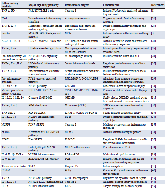
Note: TNF-α, tumor necrosis factor-alpha; IFN-γ, interferon-gamma; JAK/STAT1/IRF1, Janus kinase/signal transducer and activator of transcription 1/interferon regulatory factor 1; IL-6, interleukin-6; MSR, macrophage scavenger receptor; PAD4, peptidyl arginine deiminase 4; ROS, reactive oxygen species; NETs, neutrophil extracellular traps; ACOD1, aconitate decarboxylase 1; IRG1, immunoresponsive gene 1; CDK2, cyclin-dependent kinase 2; MAPK8, mitogen-activated protein kinase 8; JUN, Jun proto-oncogene; HIF-1α, hypoxia-inducible factor 1-alpha;
2.3 Cellular communication and inflammatory cascades
The septic cytokine storm is characterized by cell-to-cell communication, which is the driving force for systemic cascading inflammation. Communication between immune cells and endothelial cells is also key to this process. They form an intricate signaling network, connected by numerous means such as direct cellular contact, excretion of soluble factors, and extracellular vesicles (EVs), thus coordinating the initiation, amplification, and resolution of inflammation. Experiments have shown that direct interaction between bone marrow-derived mesenchymal cells and macrophages greatly enhances the production of IL-6, IL-10, and nitric oxide (NO). The generation of TNF-α is also suppressed by prostaglandin E2. As a result, bone marrow-derived mesenchymal cells may alleviate systemic inflammation and modulate tissue inflammation, thereby maintaining immune homeostasis [46]. EVs carry MFG-E8, which facilitates the complete phagocytosis of apoptotic cells and therefore decreases systemic inflammation, as well as serving a protective function in sepsis [47]. In addition, bone marrow-derived mesenchymal stem cell-derived exosomes induce M2 macrophage polarization by targeting the miR-20a-5p/CXCL12 axis, thereby effectively inhibiting the septic cascade [48].
By recruiting immune cells and regulating inflammation, Meteorin-like protein can direct the migration of macrophages, which in turn dramatically increases the body’s resistance to sepsis, even as it balances new controls over Treg/Th17 immunity [49]. However, via the Snail/CXCL2 axis, PAK1 can enhance the inflammatory cascade following sepsis [50]. It is worth noting that in both infectious and noninfectious systemic inflammation, IL-6 can promote TNF-α production through the NF-κB pathway rather than classical STAT3 signaling, thus making inflammatory responses more severe [51].
In sepsis, there exists a highly complex and tightly coupled intracellular signaling network. Through multiple pathways within these networks, the inflammatory cascade can spread rapidly. The mechanisms by which innate immune activation and inflammatory cell death contribute to sepsis are dissected in this review by summarizing the classical and non-classical activation processes of the inflammasome signaling pathway in Figure 1. Via the TLR-NF-κB pathway, pathogen-associated molecular patterns induce the expression of pro-IL-1β and NLRP3. LPS can activate non-classical pathways mediated by caspase-11; this in turn leads to formation of the NLRP3-apoptosis-associated speck-like protein containing a CARD (ASC)-caspase-1 complex, in which GSDMD is cleaved and N-terminal pores are formed. This can induce pyroptosis and promote the maturation and release of IL-1β, thereby amplifying dysregulated inflammation and aggravating organ dysfunction during sepsis. As the understanding of these communication and signaling networks becomes more profound, the etiology of the cytokine storm is revealed, providing both a theoretical basis and a strategy for precise anti-inflammatory intervention by regulating major pathways or EV release.
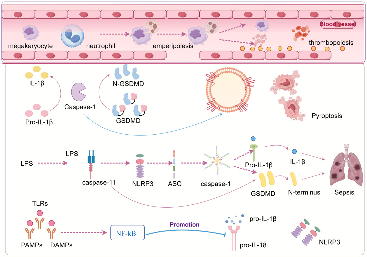
Figure 1. Mechanistic diagram of inflammasome activation and pyroptosis in sepsis and thrombopoiesis. This diagram illustrates how inflammasome activation contributes to excessive inflammatory responses and subsequent organ dysfunction during sepsis, as well as to platelet formation via GSDMD-driven pyroptosis. TLRs recognize PAMPs and DAMPs and then activate NF-κB signaling, prompting the expression of pro-inflammatory cytokines such as pro-IL-1β and pro-IL-18, as well as NLRP3. Activation of caspase-11 by LPS contributes to NLRP3 inflammasome assembly, and caspase-1 is also activated at the plasma membrane via the adaptor protein ASC. N-GSDMD can form membrane pores, thereby enabling pyroptosis as well as the release of IL-1β. Thus, in sepsis model mice, this process accelerates both systemic inflammation and disease progression. The inset graph illustrates emperipolesis occuring between neutrophils and megakaryocytes. It suggests that this specific cellular response leads to both enhanced platelet formation and amplification of inflammation in the local vascular environment.
3.1 Microcirculatory dysfunction and organ failure
Sepsis is defined as life-threatening organ dysfunction caused by a dysregulated host response to infection. Microcirculatory failure represents one of the key pathophysiological mechanisms underlying organ dysfunction in sepsis. This non-physiological state not only fails to provide acceptable tissue perfusion but also exacerbates the course of multiorgan failure [52, 53]. In sepsis, elevated PCSK9 levels can activate the TLR4/MyD88/NF-κB and NLRP3 pathways, thereby promoting dysregulated systemic inflammation and endothelial injury that contribute to organ dysfunction [54]. Concurrently, endothelial cell activation, upregulation of adhesion molecules, and increased vascular permeability enhance plasma extravasation and tissue hypoperfusion. Increased expression of Mac-1 on the neutrophil surface induces pulmonary microcirculatory dead space, and inhibitors of Mac-1 can partially improve ALI in sepsis, suggesting that microcirculatory perfusion abnormality is an important feature during organ dysfunction [55].
Microthrombosis is also a significant cause of microvascular dysfunction in sepsis. TNF-α, IL-1β, and HMGB1-associated proinflammatory signals promote both platelet aggregation and fibrin deposition, leading to subsequent capillary occlusion and non-uniform blood flow. Furthermore, high PAI-1 levels represent a disseminated intravascular coagulation state with suppressed fibrinolysis and are strongly related to organ dysfunction [53]. Reduced erythrocyte deformability and reduced blood flow worsen tissue hypoxia, such that microcirculatory perfusion may be impaired despite normal blood flow in the larger vessels, leading to a mismatch between perfusion and oxygenation even when large vessel blood flow is unimpaired [52]. Both clinical and experimental studies have demonstrated that septic shock is associated with peripheral vascular hyporesponsiveness, although some capillary beds within the post-capillary circulation retain residual responsiveness [56]. These findings imply that vasomotor regulatory function is an important factor in sustaining microcirculatory integrity.
Some interventions targeting microcirculatory failure have beneficial effects. For instance, Shenfu Injection enhances microcirculatory perfusion and endothelial function through suppression of PI3K/Akt-induced glycolysis [57]. Salvianolic acid B, meanwhile, mitigates microcirculatory failure and sepsis progression by suppressing platelet CD226 molecule function [58]. Multiple factors, including endothelial damage, an overwhelming inflammatory response, microthrombosis, and imbalanced oxygen delivery, play a role in the pathogenesis of septic microcirculatory dysfunction. These mechanisms interact to generate a complex network, resulting in multi-organ failure. This explains the clinical approach toward multi-target interventions (microcirculation protection, anti-inflammatory treatment, and antithrombosis therapy).
3.2 Pathogen recognition and host response
Sepsis is a severe syndrome caused by microbial infection and characterized by excessive host inflammatory responses and organ injury [59]. In this process, pathogen-associated molecular patterns and damage-associated molecular patterns are recognized by pattern recognition receptors expressed on epithelial and endothelial cells of barrier tissues, as well as on circulating and resident innate immune cells, thereby initiating early innate immune responses. This represents a generalized mechanism of early host–pathogen interaction in sepsis rather than a process restricted to a specific anatomical site or experimental model, thereby triggering the initial immune defense [60].
Neutrophils assemble inflammasome complexes in distinct subcellular compartments and, in sepsis-relevant models, predominantly release mature IL-1β and IL-18. In these settings, extracellular IL-1α and IL-33 are often minimal or undetectable, indicating a context-dependent and selective inflammasome-associated cytokine output rather than a universal paradigm. This suggests that the pro-inflammatory function of the inflammasome during sepsis is highly selective [60].
In monocytes, TLR agonists induce an overall similar pattern of glycoprotein expression changes, but the profile changes under Staphylococcus aureus stimulation is partially distinct. Eleven glycoproteins (CD44, CD274, LILRB1, ICAM1, DSC2, PTGS2, LAMP3, CR1, FGL2, HP, and SLC1A3) are consistently dysregulated in the tolerant cell state [61].
C-ter100 can activate NF-κB through TLR4 and promote TNF-α and NO generation, as well as provoke NLRP3 inflammasome assembly (NLRP3, ASC, caspase-1) and IL-1β secretion [62]. Loss of nucleic acid recognition is likely to contribute to the lack of early detection of S. pyogenes, diminishing local infection control and ultimately initiating systemic inflammation at later stages [63]. Expression and regulation of monocyte TLR-2 may be abnormal in septic patients, whereas those for TLR-4 are relatively low [64].
Peritonitis can increase expression of peritoneal exudate cells genes involved in sepsis but does not augment this response after LPS pretreatment [65]. FLAP and 5-LO-dependent eicosanoids are also involved in bacterial endotoxin-induced inflammation through TLR-dependent mechanisms [66]. Genetic background, gut microbiota composition, and metabolic status modulate the magnitude and kinetics of the host immune response. Together, these components of the ambivalent inflammatory response in sepsis dictate whether pathogen elimination through inflammation, or instead, characteristic hyperinflammation will prevail.
3.3 Metabolic reprogramming and cell death
Metabolic reprogramming and pyroptosis during sepsis progression are mutually intertwined, propagating the inflammatory response and ultimately resulting in multiorgan failure. It has been reported that these interventions, including scavenging of lipid peroxidation by the antioxidant vitamin E, chemical inhibition of PLCG1, and gene deletion for Caspase-11/GSDMD, could significantly modulate ferroptosis and other metabolism-dependent cell death pathways and ameliorate tissue damage induced by polymicrobial sepsis [67].
EGFR mediates the membrane translocation of Glut1 via the downstream TBK1/Exo84/RalA protein pathway, which results in Warburg effect. This reinforces the activation and apoptosis of CD4+ T lymphocytes, further resulting in damage to immune function and contributing to accelerated immunological exhaustion. Furthermore, IL-1R2 physically interacts with enolase 1, which can suppress glycolysis-mediated pyroptosis and inflammation, thus indicating a therapeutic target within this pathway. Dexmedetomidine can also help maintain metabolic balance by upregulating Nrf2, inhibiting mitochondrial fission, and downregulating ferroptosis and vascular leakage [68]. Meanwhile, aerobic glycolysis suppression can activate autophagy via the lactate/SIRT3/AMPK signaling pathway, thereby providing protection in sepsis-associated acute kidney injury (AKI) [69].
Mechanistically, ferroptosis and lipophagy contribute significantly to septic AKI, which has added new insight into the pathogenetic mechanisms and therapeutic targets of AKI [70]. Moreover, knockout or knockdown of MAPL also attenuates septic myocardial injury and inflammation by inhibiting Drp1 SUMOylation and ameliorating mitochondrial dysfunction. These findings suggest that metabolic reprogramming and multimodal cell death are tightly interconnected in sepsis and together contribute to a dysregulated host response to infection, characterized by excessive inflammation followed by immune suppression, which ultimately results in life-threatening organ dysfunctions [71].
Energy metabolism pathways can be regulated, selected forms of cell death can be inhibited, and autophagy can be stimulated, resulting in attenuation of the organ injury response and systemic inflammation. To systematically reveal the pathological processes and clinical manifestations critically associated with sepsis development, priority mechanisms are summarized in Table 2. The pathophysiology of sepsis is complex and multifaceted, comprising interactions among immune dysfunction, microcirculatory derangement, metabolic disarray, and cell death networks. These mechanisms collectively contribute to the progression from localized infection to dysregulated systemic host response and subsequent organ dysfunction: microcirculatory dysfunction induces tissue hypoxia; abnormal recognition and/or excessive response to pathogens will excessively activate the immune system; and metabolic disorders activated by these former processes, along with programmed cell death itself, further enhance inflammation-induced injury. This mechanistic framework informs the pathogenesis of sepsis and identifies core nodes with their intrinsic relationships at different levels, from molecule to system, thus offering theoretical support for early identification and multi-target combination interventions.
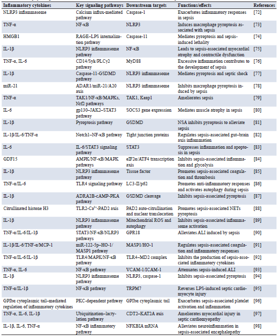
Note: NLRP3, NLR family pyrin domain containing 3; TNF-α, tumor necrosis factor-alpha; NF-κB, nuclear factor kappa-B; HMGB1, high mobility group box 1; RAGE, receptor for advanced glycation end products; LPS, lipopolysaccharide; IL-1β, interleukin-1 beta; IL-6, interleukin-6; CD14, cluster of differentiation 14; Syk, spleen tyrosine kinase; PLCγ2, phospholipase C gamma 2; MyD88, myeloid differentiation primary response 88; GSDMD, gasdermin D; miR-21, microRNA-21; ADAR1, adenosine deaminase acting on RNA 1; A20, tumor necrosis factor alphainduced protein 3 (TNFAIP3); TAK1, transforming growth factor-beta-activated kinase 1; MAPKs, mitogen-activated protein kinases; Nrf2, nuclear factor erythroid 2-related factor 2; Keap1, Kelch-like ECH-associated protein 1; gp130, glycoprotein 130; JAK2, Janus kinase 2; STAT3, signal transducer and activator of transcription 3; SOCS3, suppressor of cytokine signaling 3; NSA, necrosulfonamide; Notch1, neurogenic locus notch homolog protein 1; GDF15, growth differentiation factor 15; AMPK, AMP-activated protein kinase; eIF2α, eukaryotic initiation factor 2 alpha; ATF4, activating transcription factor 4; TLR4, Toll-like receptor 4; LC3-II, microtubule-associated protein 1 light chain 3-II; p62, sequestosome-1; ADRA2B, adrenoceptor alpha 2B; cAMP, cyclic adenosine monophosphate; PKA, protein kinase A; PAD2, peptidyl arginine deiminase 2; ROS, reactive oxygen species; GPR18, G protein-coupled receptor 18; ALI, acute lung injury; MCP-1, monocyte chemoattractant protein-1; HO-1, heme oxygenase-1; MASP1, mannan-binding lectin serine protease 1; MD2, myeloid differentiation factor 2; VCAM-1, vascular cell adhesion molecule 1; ICAM-1, intercellular adhesion molecule 1; TRPM7, transient receptor potential melastatin 7; GPIbα, glycoprotein Ib alpha chain; PKC, protein kinase C; CDT2, full name to be confirmed, a RING-type E3 ubiquitin ligase; KAT2A, lysine acetyltransferase 2A; NFKBIA, nuclear factor of kappa light polypeptide gene enhancer in B-cells inhibitor alpha.
4.1 Immunosuppressive phase and immune evasion
Studies have demonstrated that the immunosuppressive phase of sepsis involves sophisticated immune regulation and evasion, which are mediated through broad interplays among cellular functions, metabolism, and signaling. Neutrophils with high expression of CD200 receptor may also induce systemic immunosuppression by promoting Treg cells [99]. Indeed, recent investigations also indicate that metabolic reprogramming acts as a pivotal mediator of crosstalk between immune cell subsets in sepsis. Inflammatory stress and hypoxia direct immune cells into a state of increased glycolysis and modified lipid metabolism, resulting in remodeling of immune differentiation and functional polarization. Metabolic rewiring, for instance, skews T-cell subsets toward regulatory phenotypes, and lactate accumulation inhibits effector T-cell responses. Macrophage polarization is tightly associated with metabolic state. These results suggest that metabolic changes actively drive immune subset dysregulation during sepsis and are not simply a reflection of immune activation [100]. This indicates that intracellular metabolic regulation could be a novel therapeutic approach for sepsis-induced immunosuppression [101]. CXCR2+ neutrophil subtypes also participate in immune suppression, which may warrant their consideration as therapeutic targets [102].
TCF7 and LEF-1 overexpression can significantly upregulate the proliferation capability and effector function of CD4+ T cells, inhibit sepsis-induced apoptosis, and downregulate the expression of PD-1/LAG-3, resulting in a stronger immune response in patients with sepsis [103]. Metabolic dysfunctions have an intrinsic role in immunosuppression: lactate impairs T cell activation by downregulating CD40LG and SOCS3 expression, consequently restraining the JAK-STAT signaling pathway [104]. Low-density neutrophils suppress T cells through PD-L1 and contribute to susceptibility to secondary infections [105]. Neutrophil immune function can be compromised by glycolysis blockage, which downregulates lactate dehydrogenase A via the PI3K/Akt-HIF-1α signaling pathway [106]. The Spns2/S1P signaling pathway is important for regulating immune homeostasis, inhibiting excessive early inflammation, and relieving delayed immunosuppression, which may have a critical role in immune remodeling [107].
These investigations systematically demonstrate the hallmarks of regulatory networks in the immunosuppressive process of sepsis and underscore that the generation of Treg cells, subset properties of neutrophils, and dysregulated cellular metabolism are crucial hubs for immune evasion. They support rational drug discovery targeting metabolic reprogramming, immune checkpoint regulation, and signaling pathways.
4.2 Negative feedback mechanisms and host homeostasis
Targeted deletion of IL-10 in CD169+ macrophages results in dramatically increased mortality during septic challenge, and administration of recombinant IL-10 is protective in a model of LPS-induced lethality, thereby highlighting the importance of anti-inflammatory mediators during early negative feedback regulation of inflammation [108, 109]. Adenosine deaminase acting on RNA 1 (ADAR1) inhibits sepsis-related ALI by suppressing the activation of pyroptosis in pulmonary macrophages via the miR-21/A20/NLRP3 axis, demonstrating that the host provides a possible negative feedback-based control mechanism to prevent inflammation propagation and tissue damage at the molecular level [78]. In patients with sepsis, T cell immunoreceptor with Ig and ITIM domains (TIGIT)+ T cells display PD-1 upregulation, CD226 downregulation, and impaired cytokine secretion, remarkably, in vitro blocking of TIGIT restores T cell activity. These results imply that the inhibitory receptor modulates T cell activity through negative signaling to balance the immune response and avoid excessive inflammation [110].
The IL-10/DEL-1 axis not only favors emergency granulopoiesis over neutropenia but also promotes early host survival by balancing the abundance and function of immune cells, thus emphasizing the role of immune negative feedback in inducing overall immune homeostasis [108, 109, 111]. In addition, the finding that the absence of IL-10 in B cells leads to aberrant cGMP-PKG signaling, and that exogenous IL-10 supplementation alleviates LPS-induced ALI, suggests that negative feedback loops maintain both local and systemic immune homeostasis through dense network interactions with cytokines and immune cells [111]. In CLP-induced sepsis survivor mice, splenic CD11b+ Ly6Chigh myeloid cells are significantly expanded at 4 weeks post-injury, and are predominantly composed of monocytic myeloid-derived suppressor cells with evident metabolic reprogramming. Overall, this population exhibits M-MDSC–like metabolic features, suggesting that immunosuppressive myeloid cells participate in post-inflammatory immune regulation after sepsis in a dynamic and adaptive manner [112].
IL-4 induces LAMP2 expression by activating STAT3 in lysosomal homeostasis and autophagic flux, and this metabolic and cellular homeostatic regulation is also involved in negative feedback control [113]. Furthermore, artemisinin-based therapy rescues both inflammatory and immunosuppressive states in sepsis immune cells, therefore globally restoring the homeostasis of the host’s immunological defense function, suggesting that these negative feedback mechanisms play an integrative regulatory role in controlling host defense.
4.3 Role of immunoregulatory cells
During the development of sepsis and the cytokine storm, immunoregulatory cells are critically involved in the regulation of host immune homeostasis and the downregulation of excessive inflammation. They inhibit hyperactivation of effector T cells, dendritic cells, and other immune-activated cells through the growth factors IL-10 and TGF-β, as well as through contact-mediated inhibitory mechanisms, thus locally restricting inflammation to control tissue damage. Production of reactive oxygen species and NO by MDSCs is the most common mechanism to inhibit T cell proliferation, as well as to control amino acid metabolism pathways and the inflammatory activity of macrophages and neutrophils. At the height of inflammation, the explosive accumulation of MDSCs acts to protect tissues from injury, while their protracted survival can contribute to immunosuppression.
Local immune self-regulation, however, is still modulated by tissue-resident regulatory macrophages and dendritic cell subtypes that not only produce anti-inflammatory/proresolving mediators and phagocytose apoptotic cells but also regulate the expression of co-stimulatory molecules to dampen hyperresponsiveness, thereby essentially inhibiting overt inflammatory responses. At the molecular level, Nrf2 plays a role in protecting against sepsis-induced lung injury by modulating autophagy and NF-κB/PPARγ-dependent macrophage polarization [114]. GPR174 affects early immune regulation in sepsis by altering macrophage phenotype and the production of both pro- and anti-inflammatory cytokines [10]. In addition, the ‘mature DCs enriched in immunoregulatory molecules’ program is activated within 24 hours after sepsis onset via the TNFRSF-NF-κB and IFNGR2-JAK-STAT3 pathways, highlighting the critical role of dendritic cells in early immune modulation [115].
PAD2/PAD4 deletion restrains NLRP3 activation and accelerates the resolution of inflammation by skewing the Ym1+ M2 macrophage phenotype toward a resolving phenotype in the lungs. Thus, the PADIs/NLRP3/Ym1 axis may be a novel therapeutic target for sepsis-associated ALI [116]. Repeated sepsis worsens CD4+ T cell exhaustion and the antiviral immune response, leading to poor outcomes; administration of anti-TIGIT monoclonal antibodies can reverse T cell apoptosis caused by sepsis and significantly improve survival [117, 118].
Overall, dynamic regulation of Tregs, MDSCs, regulatory macrophages, and dendritic cells, combined with the factors involved in these key signaling pathways, constitutes a complicated immunoregulatory network in sepsis and provides an excellent theoretical basis for precision intervention and targeted treatment.
5.1 Precision medicine and personalized therapy
BAM15 promotes mitochondrial DNA-dependent responses and may serve as a potential companion biomarker for the initial diagnosis and efficacy monitoring of septic patients during therapy [119]. The immune response in sepsis is highly variable, and this heterogeneity has a profound impact on disease outcome and its treatment. Integrated analysis of patients’ immune phenotypes, metabolic status, microbiome information, and genetic content leads to accurate stratification of the intensity and type of inflammatory responses and provides a solid scientific foundation for personalized interventions [120]. A typical application scenario may involve the patient population with a low resistance program molecular fingerprint compared with systemic inflammation levels, for which greater diversity of immune states is relevant for developing more sophisticated stratification strategies and adjustment of therapeutic schedules.
Certain combinations of IFN-γ and IL-1β can efficiently differentiate cytokine release syndrome from sepsis, thus significantly enhancing the accuracy of individualized therapeutic interventions [121]. Neutrophil-derived C1q has been proposed as a consistent prognostic biomarker for sepsis-associated death and could be a therapeutic target [122]. Furthermore, the transfer of apoptotic cells to patients with mild-to-moderate sepsis has been found to be safe and feasible, capable of directing immune responses and contributing to early termination of the cytokine storm [123]. As an essential autophagy-regulatory molecule, RAS protein activator-like 3 not only plays a role in the treatment of sepsis but also provides a new therapeutic target for other inflammatory conditions [124].
Using precise risk factors, such as the Acute Physiology and Chronic Health Evaluation III score, bicarbonate levels, anion gap, and invasive/noninvasive systolic blood pressure in combination, more personalized therapies can be tailored to improve survival time and prognostic outcomes in patients with sepsis-induced acute respiratory distress syndrome [125]. Furthermore, the heterogeneity of sepsis subtypes and differences in mortality among populations have been observed; such a tri-variable model can effectively identify patients with the δ sepsis subtype and thus form a reliable basis for dynamic monitoring and stratified intervention [126]. Integration of high-throughput multi-omics techniques, single-cell transcriptomic profiling, and AI algorithms allows real-time monitoring of immune cell subsets and inflammatory networks. This holistic approach can offer an evidence‐based strategy for appropriate drug choice, medication dosage, and timing of intervention in the cytokine storm to maximize its control, reconstitute immune balance, and ultimately improve patient outcomes.
5.2 Application of biomarkers in clinical decision-making
However, no single biomarker can fully capture the complexity of the inflammatory network in sepsis, further supporting the need for phenotype-oriented multimarker integration. Multimarker composite features—such as lactate, cytokine profiles, immune cell functions, and metabolism—can offer a more accurate determination of patients’ inflammatory status and immune competence for early intervention and personalized therapy. Continuous monitoring of important inflammatory mediators and immune parameters allows closer guidance of therapy with anti-infective and immunomodulatory drugs in real time, optimizing therapy duration and avoiding over-immunosuppression or overactivation. In an era of progressive development in single-cell analysis, metabolomics, and AI-driven predictive models, the clinical value of these biomarkers is anticipated to achieve a higher level of precision and personalization, providing firm support for accurate patient stratification and identification, tailored interventions, and improved outcomes in sepsis, and ultimately reducing sepsis-associated mortality.
5.3 Novel immunomodulators and anti-inflammatory agents
At the same time, the translational relevance of immunomodulatory spectra in sepsis becomes more substantive when treatment is specifically targeted to biologically delineated immune endotypes. Interventions such as adjunctive corticosteroids, rather than being applied uniformly, may be more properly considered within gene-expression-informed inflammatory states, which can identify patients likely to benefit most and mitigate the potential harms of indiscriminate immune suppression [136]. Sepsis and its attendant cytokine storm represent a condition of intricate immunopathobiology, characterized by marked dysfunction and dysregulation of the host immune response, including excessive activation of inflammation in conjunction with immunosuppression and multiple organ failure. In the context of increasingly deeper integration of omics technologies and AI, the therapeutic concept of sepsis has also been transforming from traditional anti-infection or supportive care to one involving precision stratification, dynamic monitoring, and multidimensional intervention. The multi-targeted regulation modes of immune disturbance, metabolic dysfunction, and inflammatory overreactions present challenges for patient therapy. In summary, to systematically summarize recent developments and prospects in sepsis treatment, this review addresses emerging interventional strategies reported in the past decade in Table 3, including precision medicine, biomarker-guided management, and immunomodulatory therapy, which may provide theoretical support for a personalized medicine-oriented individualized therapeutic approach as well as combined multi-target interventions.
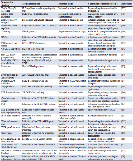
Note: Nrf2, nuclear factor erythroid 2-related factor 2; NLRP3, NLR family pyrin domain containing 3; IL-1β, interleukin-1 beta; EphA4, ephrin type-A receptor 4; Fc, fragment crystallizable; HO-1, heme oxygenase-1; NF-κB, nuclear factor kappa-B; UDCA, ursodeoxycholic acid; STING, stimulator of interferon genes; PANoptosis, pyroptosis-apoptosis-necroptosis; FRC, fibroblastic reticular cell; CD5L, CD5 molecule-like; PINK1, PTEN-induced putative kinase 1; CXCR1/2, C-X-C chemokine receptor type 1/2; NETosis, neutrophil extracellular trap formation; miR-223, microRNA-223; MALAT1, metastasis-associated lung adenocarcinoma transcript 1; STAT3, signal transducer and activator of transcription 3; p-STAT3, phosphorylated STAT3; Notch1, neurogenic locus notch homolog protein 1; AKT, protein kinase B; GSK3β, glycogen synthase kinase 3 beta; Fyn, Fyn proto-oncogene; NRF2, nuclear factor erythroid 2-related factor 2; NLRP6, NLR family pyrin domain containing 6; TRIM21, tripartite motif-containing protein 21; TAB1, TGF-beta-activated kinase 1 binding protein 1; PI3K, phosphoinositide 3-kinase; Fgr, Gardner-Rasheed feline sarcoma viral oncogene homolog; SIRT1, sirtuin 1; PGC-1α, peroxisome proliferator-activated receptor gamma coactivator 1-alpha; FGF1, fibroblast growth factor 1; IL-6, interleukin-6; ELT, esculetin; ALI, acute lung injury; IL-40, interleukin-40; S100A8/9, S100 calcium-binding protein A8/A9; ZBP1, Z-DNA binding protein 1; TIFA, TRAF-interacting protein with forkhead-associated domain; MSM, methylsulfonylmethane; H3K18la, histone H3 lysine 18 lactylation; Arg1, arginase 1; VDR, vitamin D receptor; FFAR2, free fatty acid receptor 2; ATP, adenosine triphosphate; TAK1, transforming growth factor-beta-activated kinase 1; MAPKs, mitogen-activated protein kinases; CLP, cecal ligation and puncture.
In addition, melittin was demonstrated to relieve sepsis-induced AKI by suppressing ferroptosis mediated by the GPX4/NRF2 signaling [159]. GDF15 protects against sepsis-induced pulmonary injury by activating AMPK, inhibiting glycolysis, and inactivating the NF-κB/MAPK signaling pathways, leading to anti-inflammatory polarization of alveolar macrophages [84]. NWG specifically inhibits Src, AKT1, and cyclooxygenase-2 (COX-2) to suppress the Src/AKT1/NF-κB signaling pathway, resulting in anti-inflammatory effects and protection of lung microvascular barrier function [160]. In murine models of acute respiratory distress syndrome and sepsis, Dex@GNPs with natural glycyrrhizin protein nanoparticles can remodel the disorganized immune microenvironment and reduce tissue damage, presenting an effective method for localized anti-inflammatory and immunity regulation [161]. Dragon’s blood pigment, a CMPK2 blocker, may be therapeutically effective in sepsis by regulating metabolic and signaling pathways [162]. Enoxolone-mediated sepsis suppression involving the NF-κB pathway and MEK/ERK signaling pathways suggests a new therapeutic approach that combines precise control of cellular signal transduction with anti-inflammatory treatment strategies [163]. Furthermore, miR-223 overexpression in macrophages hinders their polarization toward the M1 phenotype after LPS stimulation and decreases sepsis severity in the context of IL-4 pretreatment. These findings provide new insight into the induction of anti-inflammatory macrophages via regulation of cellular energy metabolism and provide a basis for macrophage-based cell therapy in sepsis [144].
The balance between immune activation and immunosuppression also depends on the pathogenesis of sepsis, and immunomodulation is the main therapeutic direction in infected patients. Here, recent major treatment strategies for sepsis are systematically characterized: immune checkpoint blockade, cellular and cytokine interventions, and targeted drug-delivery systems in Figure 2. In particular, PD-1/PD-L1 and cytotoxic T-lymphocyte-associated protein 4 (CTLA-4) inhibitors reverse immunosuppression and restore T cell effector function; thymosin α1 and cell-based products enhance host immune defense; GM-CSF and IFN-γ alleviate immune paralysis; and the modulation of macrophage M1/M2 polarization via pharmacological agents or nanoparticle delivery systems is involved in the precision treatment of the inflammatory response. Collectively, these strategies achieve targeted regulation of key nodes within the inflammatory network, suppressing excessive inflammation while preserving host defense mechanisms and multiorgan homeostasis, thereby providing novel theoretical frameworks and practical pathways for multi-level, controllable intervention in sepsis. While novel immunomodulatory therapies (melittin, GDF15, NWG, 4-OI, and nanoparticle-mediated delivery systems) show great promise in animal models of sepsis/sepsis-associated encephalopathy as well as in their in vitro counterpart, there is still a lack of clinical validation in patients, and few have been validated in national cohort studies with large sample sizes suggesting high specific efficacy. Specifically, the diagnostic value of GDF15 and its potential as a therapeutic target in sepsis-associated encephalopathy merit further experimental and clinical studies [164]. Furthermore, safety issues, dose optimization, pharmacokinetics, and patient heterogeneity require thorough evaluation in well-designed clinical trials. As such, these approaches should currently be considered promising therapeutic candidates rather than established clinical options.
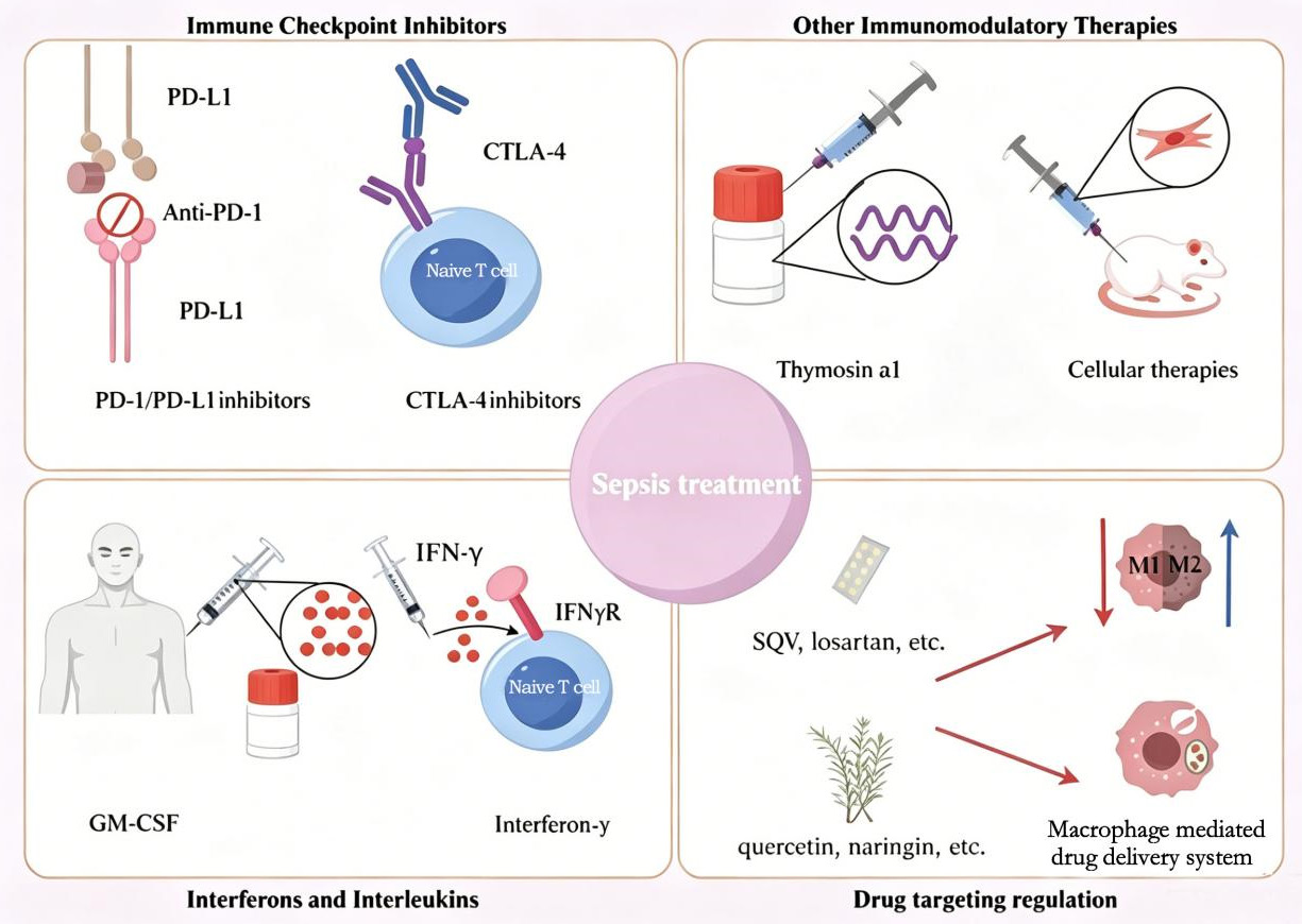
Figure 2. Immunomodulation and targeted intervention in sepsis. This figure shows the main immunomodulatory and targeted therapy strategies for treating sepsis. Anti-PD-1/PD-L1 and CTLA-4 blocking antibodies, as immune checkpoint inhibitors, can restore T cell function and ameliorate post-septic immunosuppression. Other immunomodulatory treatments, such as thymosin α1 or cellular therapy, also improve host immune defense and tissue recovery. Interferons and interleukins, such as GM-CSF and IFN-γ, promote immune cell activation and alleviate immune dysfunction. In drug-targeted regulatory strategies, the use of pharmacological agents (such as SQV, losartan, and quercetin), naringin, or macrophage-based drug delivery systems to regulate the balance between M1/M2 polarization can fine-tune the induction of inflammatory responses and favor the resolution of immune homeostasis re-establishment.
This review comprehensively demonstrates the complex network properties of sepsis and the cytokine storm, including immune hyperactivation, cytokine networks, intercellular signaling crosstalk, microcirculatory dysfunction, metabolic remodeling, and multimodal cell death, revealing their multidirectional pathological mechanisms. As the core mechanism, the cytokine storm not only accelerates multiple organ dysfunction but also further promotes immune imbalance through a positive-feedback mechanism, thereby limiting the effectiveness of single-target interventions. Inhibition of the expression or activity of DNA-PKcs may be a promising strategy for treating sepsis, as it may inhibit or mitigate mitochondrial dysfunction and organ damage in sepsis-related MODS.
Current treatment for sepsis is mainly limited to anti-infective therapy, fluid resuscitation, and organ support. However, these strategies focus one-sidedly on the symptoms and complications of the disease, which is insufficient to regulate the complicated inflammatory process. The pathophysiology of sepsis consists of signaling networks and metabolic systems interacting at multiple levels, and its nonlinear and dynamic nature suggests that single interventions may be insufficient to entirely suppress inflammatory cascades or return the immune system to homeostasis. Thus, multi-target and controllable interventions have been considered an important future direction. For instance, 4-OI has multi-target protective effects on LPS-induced septic AKI by inhibiting the inflammatory response and oxidative stress as well as promoting mitophagy [165].
Biomarkers and precision medicine hold promise in sepsis care. Early identification of high-risk patients, dynamic tracking of inflammation status, and incorporation of combined features of cytokine signatures, immune cell function, and metabolism may open options for stratified interventions and provide a support system for precise clinical decisions. The presence of different sepsis subphenotypes, as well as their relationship to disease trajectory, clinical presentations, and outcomes, has been identified through large-scale proteomics studies, leading the way to new biomarkers and precision-based interventions.
New immunomodulatory agents and anti-inflammatory interventions also offer intriguing treatment options for sepsis. For example, NAD(H)-loaded nanoparticles can enhance energy supply and inhibit inflammation, as well as promote immune homeostasis and vascular function [166]. Obeticholic acid also suppresses pro-inflammatory mediator synthesis and mitochondrial damage by reducing oxidative stress through inhibition of the NF-κB/NLRP3 signaling pathway [167]. Taken together, these results argue for a paradigm shift in sepsis therapy, incorporating multi-level, dynamically controlled network intervention paradigms and moving away from rigid single-target approaches.
However, translational efforts and clinical practice still encounter several difficulties, such as the large heterogeneity of inflammatory networks, individual variability in immunological responses, and limitations of existing biomarkers. Notably, the combination of high-throughput omics technologies, single-cell analyses, and AI-based predictive models is anticipated to enhance early risk stratification and precision intervention, as well as to provide a theoretical basis for multi-target combination therapies [168]. Importantly, a study that utilized the Surviving Sepsis Campaign guidelines and applied machine learning to integrate six major treatments—including antibacterial therapy, balanced crystalloids, insulin therapy, corticosteroids, vasopressin, and bicarbonate—into a bundled algorithm, which significantly decreased 28-day mortality in patients with sepsis and septic shock. These findings indicate that, in the future, improving outcomes will be achieved predominantly through integrating network-based multidimensional interventions with precision medicine strategies.
4-OI, 4-octyl itaconate; AKI, acute kidney injury; ALI, acute lung injury; ASC, apoptosis-associated speck-like protein containing a CARD; BMP9, bone morphogenetic protein 9; COX-2, cyclooxygenase-2; CTLA-4, cytotoxic T-lymphocyte-associated protein 4; EGFR, epidermal growth factor receptor; ERK, extracellular signal-regulated kinase; EV, extracellular vesicle; GM-CSF, granulocyte-macrophage colony-stimulating factor; GSDMD, gasdermin D; IDO1, indoleamine 2,3-dioxygenase 1; LPS, lipopolysaccharide; Mac-1, macrophage-1 antigen; MAPK, mitogen-activated protein kinase; MDSC, myeloid-derived suppressor cell; MODS, multiple organ dysfunction syndrome; MyD88, myeloid differentiation primary response 88; NET, neutrophil extracellular trap; NLRP3, NOD-like receptor family pyrin domain containing 3; NO, nitric oxide; NWG, N-(1,3,4-oxadiazol-2-yl) guanidine; PTX3, pentraxin 3; SOFA, sequential organ failure assessment; TIGIT, T cell immunoreceptor with Ig and ITIM domains; TLR, Toll-like receptor; Treg, regulatory T cell.
Author contributions
Linzhu Li and Kaikai Wang contributed equally to this work and share first authorship. Linzhu Li and Kaikai Wang were responsible for literature retrieval and manuscript drafting. Yongbo Li participated in the design of the article framework and manuscript revision. Wenrong Zhang, Jianlong Ma, and Wenzhi Zhang were responsible for literature screening, summarizing research progress, and content organization. Wanquan Guo and Qianqian Zhang participated in literature analysis, figure design, and language polishing. Zhijing Song was responsible for the overall study design, academic supervision, manuscript review, and final approval of the manuscript. All authors have read and approved the final version of the manuscript.
Funding
This research received no external funding.
Data availability
Data sharing is not applicable to this article as no new data were created or analyzed in this study.
Ethics approval and consent to participate
Not applicable.
Consent for publication
Not applicable.
Competing interests
The authors declare no conflict of interest.
Acknowledgements
Not applicable.
[1] Limmer A, Engler A, Kattner S, Gregorius J, Pattberg KT, Schulz R, et al. Patients with SARS-CoV-2-induced viral sepsis simultaneously show immune activation, impaired immune function and a procoagulatory disease state. Vaccines (Basel). 2023 Feb 13;11(2):435. https://doi.org/10.3390/vaccines11020435
[2] Rudd KE, Johnson SC, Agesa KM, Shackelford KA, Tsoi D, Kievlan DR, et al. Global, regional, and national sepsis incidence and mortality, 1990-2017: Analysis for the global burden of disease study. Lancet. 2020 Jan 18;395(10219):200-211. https://doi.org/10.1016/s0140-6736(19)32989-7
[3] Zhang H, Wu D, Wang Y, Shi Y, Shao Y, Zeng F, et al. Ferritin-mediated neutrophil extracellular traps formation and cytokine storm via macrophage scavenger receptor in sepsis-associated lung injury. Cell Commun Signal. 2024 Feb 2;22(1):97. https://doi.org/10.1186/s12964-023-01440-6
[4] Hou J, Zhang J, Cui P, Zhou Y, Liu C, Wu X, et al. TREM2 sustains macrophage-hepatocyte metabolic coordination in nonalcoholic fatty liver disease and sepsis. J Clin Invest. 2021 Feb 15;131(4):e135197. https://doi.org/10.1172/jci135197
[5] Cheng MP, Stenstrom R, Paquette K, Stabler SN, Akhter M, Davidson AC, et al. Blood culture results before and after antimicrobial administration in patients with severe manifestations of sepsis: A diagnostic study. Ann Intern Med. 2019 Oct 15;171(8):547-554. https://doi.org/10.7326/m19-1696
[6] Niu Y, Xu G, Zhu S, Wei X, Wu C, Zhang R, et al. NONO regulates multiple cytokine production in sepsis via the ERK1/2 signaling pathway. Mol Immunol. 2023 Jan;153:94-105. https://doi.org/10.1016/j.molimm.2022.11.017
[7] Lin L, Xu L, Lv W, Han L, Xiang Y, Fu L, et al. An NLRP3 inflammasome-triggered cytokine storm contributes to Streptococcal toxic shock-like syndrome (STSLS). PLOS Pathog. 2019 Jun 6;15(6):e1007795. https://doi.org/10.1371/journal.ppat.1007795
[8] Karki R, Sharma BR, Tuladhar S, Williams EP, Zalduondo L, Samir P, et al. Synergism of TNF-α and IFN-γ triggers inflammatory cell death, tissue damage, and mortality in SARS-CoV-2 infection and cytokine shock syndromes. Cell. 2021 Jan 7;184(1):149-168.e117. https://doi.org/10.1016/j.cell.2020.11.025
[9] Fang J, Ding H, Huang J, Liu W, Hong T, Yang J, et al. Mac-1 blockade impedes adhesion-dependent neutrophil extracellular trap formation and ameliorates lung injury in LPS-induced sepsis. Front Immunol. 2025 Mar 28;16:1548913. https://doi.org/10.3389/fimmu.2025.1548913
[10] Qiu D, Chu X, Hua L, Yang Y, Li K, Han Y, et al. Gpr174-deficient regulatory T cells decrease cytokine storm in septic mice. Cell Death Dis. 2019 Mar 8;10(3):233. https://doi.org/10.1038/s41419-019-1462-z
[11] Supino D, Davoudian S, Silva-Gomes R, Piovani D, Garuti R, Desai A, et al. Monocyte-macrophage membrane expression of IL-1R2 is a severity biomarker in sepsis. Cell Death Dis. 2025 Apr 10;16(1):269. https://doi.org/10.1038/s41419-025-07597-x
[12] Chen Y, Ying J, Li Z, He Z, Zhan J, Liang H, et al. IDO1 inhibitors block septic cytokine storm by suppressing the IDO1-AHR-CYP1A1 axis. Biomed Pharmacother. 2025 Jun;187:118054. https://doi.org/10.1016/j.biopha.2025.118054
[13] Zhang F, Guo F, Zhang Y, Xu H, Liu Y, Lin L, et al. Huashibaidu formula attenuates sepsis-induced acute lung injury via suppressing cytokine storm: Implications for treatment of COVID-19. Phytomedicine. 2023 Jan;109:154549. https://doi.org/10.1016/j.phymed.2022.154549
[14] Kang S, Tanaka T, Inoue H, Ono C, Hashimoto S, Kioi Y, et al. IL-6 trans-signaling induces plasminogen activator inhibitor-1 from vascular endothelial cells in cytokine release syndrome. Proc Natl Acad Sci U S A. 2020 Sep 8;117(36):22351-22356. https://doi.org/10.1073/pnas.2010229117
[15] Li W, Li Y, Qin K, Du B, Li T, Yuan H, et al. Siglec-G deficiency ameliorates hyper-inflammation and immune collapse in sepsis via regulating Src activation. Front Immunol. 2019 Nov 7;10:2575. https://doi.org/10.3389/fimmu.2019.02575
[16] Verweyen E, Holzinger D, Weinhage T, Hinze C, Wittkowski H, Pickkers P, et al. Synergistic signaling of TLR and IFN-β facilitates escape of IL-18 expression from endotoxin tolerance. Am J Respir Crit Care Med. 2020 Mar 1;201(5):526-539. https://doi.org/10.1164/rccm.201903-0659OC
[17] Wang H, Lan Y, Luo L, Xiao Y, Meng X, Zeng Y, et al. The Scutellaria-Coptis herb couple and its active small-molecule ingredient wagonoside alleviate cytokine storm by regulating the CD39/NLRP3/GSDMD signaling pathway. J Ethnopharmacol. 2025 Jan 10;336:118741. https://doi.org/10.1016/j.jep.2024.118741
[18] Liang S, Zhou J, Cao C, Liu Y, Ming S, Liu X, et al. GITR exacerbates lysophosphatidylcholine-induced macrophage pyroptosis in sepsis via posttranslational regulation of NLRP3. Cell Mol Immunol. 2024 Jul;21(7):674-688. https://doi.org/10.1038/s41423-024-01170-w
[19] Lu J, Hou Y, Liu S, Jin B, Liu J, Li N, et al. Acetyl-CoA synthetase 2 induces pyroptosis and inflammation of renal epithelial tubular cells in sepsis-induced acute kidney injury by upregulating the KLF5/NF-κB pathway. Cell Commun Signal. 2024 Mar 21;22(1):187. https://doi.org/10.1186/s12964-024-01556-3
[20] Haschka D, Tymoszuk P, Petzer V, Hilbe R, Heeke S, Dichtl S, et al. Ferritin H deficiency deteriorates cellular iron handling and worsens Salmonella typhimurium infection by triggering hyperinflammation. JCI Insight. 2021 Jul 8;6(13):e141760. https://doi.org/10.1172/jci.insight.141760
[21] Zhang H, Zhang X, Ling C, Liu C, Hua S, Xiong Z, et al. EGFR-TNFR1 pathway in endothelial cell facilitates acute lung injury by NF-κB/MAPK-mediated inflammation and RIP3-dependent necroptosis. Int Immunopharmacol. 2023 Apr;117:109902. https://doi.org/10.1016/j.intimp.2023.109902
[22] Jung E, Romero R, Yeo L, Diaz-Primera R, Marin-Concha J, Para R, et al. The fetal inflammatory response syndrome: The origins of a concept, pathophysiology, diagnosis, and obstetrical implications. Semin Fetal Neonatal Med. 2020 Aug;25(4):101146. https://doi.org/10.1016/j.siny.2020.101146
[23] Wang D, Wang K, Liu Q, Liu M, Zhang G, Feng K, et al. A novel drug candidate for sepsis targeting heparanase by inhibiting cytokine storm. Adv Sci (Weinh). 2024 Aug;11(29):e2403337. https://doi.org/10.1002/advs.202403337
[24] Wu R, Liu J, Wang N, Zeng L, Yu C, Chen F, et al. Aconitate decarboxylase 1 is a mediator of polymicrobial sepsis. Sci Transl Med. 2022 Aug 24;14(659):eabo2028. https://doi.org/10.1126/scitranslmed.abo2028
[25] Li N, Gong Y, Zhu Y, Li B, Wang C, Wang Z, et al. Exogenous acetate attenuates inflammatory responses through HIF-1α-dependent glycolysis regulation in macrophage. Cell Mol Life Sci. 2024 Dec 27;82(1):21. https://doi.org/10.1007/s00018-024-05521-8
[26] Zheng X, Xing Y, Sun K, Jin H, Zhao W, Yu F. Combination therapy with resveratrol and celastrol using folic acid-functionalized exosomes enhances the therapeutic efficacy of sepsis. Adv Healthc Mater. 2023 Nov;12(29):e2301325. https://doi.org/10.1002/adhm.202301325
[27] Büyükcavlak M, Duman I, Eryavuz OD, Ünlü A, Duman A. Effects of artemisinin and hydroxychloroquine on cytokines in experimental sepsis. Trop Biomed. 2022 Dec 1;39(4):547-551. https://doi.org/10.47665/tb.39.4.010
[28] Eygi E, Kucuk O, Aydemir S, Atilgan M, Dokuyucu R, Erbas O. Hydroxychloroquine mitigates cytokine storm and prevents critical illness neuromyopathy in a rat sepsis model. Medicina (Kaunas). 2024 Nov 1;60(11):1791. https://doi.org/10.3390/medicina60111791
[29] Arunachalam AR, Samuel SS, Mani A, Maynard JP, Stayer KM, Dybbro E, et al. P2Y2 purinergic receptor gene deletion protects mice from bacterial endotoxin and sepsis-associated liver injury and mortality. Am J Physiol Gastrointest Liver Physiol. 2023 Nov 1;325(5):G471-G491. https://doi.org/10.1152/ajpgi.00090.2023
[30] Zou S, Han X, Luo S, Tan Q, Huang H, Yao Z, et al. Bay-117082 treats sepsis by inhibiting neutrophil extracellular traps (NETs) formation through down-regulating NLRP3/N-GSDMD. Int Immunopharmacol. 2024 Nov 15;141:112805. https://doi.org/10.1016/j.intimp.2024.112805
[31] Iske J, El Fatimy R, Nian Y, Ghouzlani A, Eskandari SK, Cetina Biefer HR, et al. NAD(+) prevents septic shock-induced death by non-canonical inflammasome blockade and IL-10 cytokine production in macrophages. Elife. 2024 Feb 19;12:RPA8866. https://doi.org/10.7554/eLife.88686
[32] Huang Y, Li G, Chen Z, Chen M, Zhai W, Li D, et al. Exosomal drug delivery systems: A novel therapy targeting PD-1 in septic-ALI. Stem Cell Rev Rep. 2024 Nov;20(8):2253-2267. https://doi.org/10.1007/s12015-024-10784-6
[33] Kuang X, Niu Z, Huang Z, Cai X, Wang L, Zhang Y, et al. GDF15 attenuates sepsis-induced acute lung injury by suppressing the HIF-1α/LDHA pathway. Int Immunopharmacol. 2025 Oct 10;163:115198. https://doi.org/10.1016/j.intimp.2025.115198
[34] Potere N, Garrad E, Kanthi Y, Di Nisio M, Kaplanski G, Bonaventura A, et al. NLRP3 inflammasome and interleukin-1 contributions to COVID-19-associated coagulopathy and immunothrombosis. Cardiovasc Res. 2023 Sep 5;119(11):2046-2060. https://doi.org/10.1093/cvr/cvad084
[35] Lan Y, Wang H, Jing L, Li R, Sun J, Meng X, et al. Jatrorrhizine alleviates cytokine storm secondary lung injury via regulating CD39-dominant purinergic braking and downstream NLRP3 inflammasome. Phytother Res. 2025 May;39(5):2374-2392. https://doi.org/10.1002/ptr.8062
[36] Xi W, Wu W, Zhou L, Zhang Q, Yang S, Huang L, et al. Multifunctional nanoparticles confers both multiple inflammatory mediators scavenging and macrophage polarization for sepsis therapy. Mater Today Bio. 2025 Feb;30:101421. https://doi.org/10.1016/j.mtbio.2024.101421
[37] Jiang T, Peng D, Shi W, Guo J, Huo S, Men L, et al. IL-6/STAT3 signaling promotes cardiac dysfunction by upregulating FUNDC1-dependent mitochondria-associated endoplasmic reticulum membranes formation in sepsis mice. Front Cardiovasc Med. 2021 Jan 18;8:790612. https://doi.org/10.3389/fcvm.2021.790612
[38] Alves GF, Aimaretti E, Einaudi G, Mastrocola R, de Oliveira JG, Collotta D, et al. Pharmacological inhibition of FAK-Pyk2 pathway protects against organ damage and prolongs the survival of septic mice. Front Immunol. 2022 Feb 1;13:837180. https://doi.org/10.3389/fimmu.2022.837180
[39] Liang X, Li H, Zhang Y, Ding Z. The flavonoids of Rosa roxburghii Tratt alleviate endotoxemia progression by inhibiting the proinflammatory effects of neutrophil and macrophage via ROS/NLRP3 signaling pathway. Int Immunopharmacol. 2025 Sep 23;162:115159. https://doi.org/10.1016/j.intimp.2025.115159
[40] Kim KM, Kim SY, Mony TJ, Bae HJ, Han SD, Lee ES, et al. Dracocephalum moldavica ethanol extract suppresses LPS-induced inflammatory responses through inhibition of the JNK/ERK/NF-κB signaling pathway and IL-6 production in RAW 264.7 macrophages and in endotoxic-treated mice. Nutrients. 2021 Dec 16;13(12):4501. https://doi.org/10.3390/nu13124501
[41] Haderski GJ, Kandar BM, Brackett CM, Toshkov IM, Johnson CP, Paszkiewicz GM, et al. TLR5 agonist entolimod reduces the adverse toxicity of TNF while preserving its antitumor effects. PLoS One. 2020 Feb 6;15(2):e0227940. https://doi.org/10.1371/journal.pone.0227940
[42] Lee SY, Hsin LW, Su MJ, ChangChien CC, Ku HC. A novel isoquinoline derivative exhibits anti-inflammatory properties and improves the outcomes of endotoxemia. Pharmacol Rep. 2019 Dec;71(6):1281-1288. https://doi.org/10.1016/j.pharep.2019.06.015
[43] Pak S, Thapa B, Lee K. Decursinol angelate mitigates sepsis induced by methicillin-resistant Staphylococcus aureus infection by modulating the inflammatory responses of macrophages. Int J Mol Sci. 2021 Oct 11;22(20):10950. https://doi.org/10.3390/ijms222010950
[44] Liu L, Lin L, Wang Y, Yan X, Li R, He M, et al. L-AP alleviates liver injury in septic mice by inhibiting macrophage activation via suppressing NF-κB and NLRP3 inflammasome/caspase-1 signal pathways. J Agric Food Chem. 2024 Apr 17;72(15):8460-8475. https://doi.org/10.1021/acs.jafc.3c02781
[45] Mukherjee D, Satyavolu S, Thomas A, Cioffi S, Li Y, Chan ER, et al. Neutrophil KLF2 regulates inflammasome-dependent neonatal mortality from endotoxemia. J Leukoc Biol. 2025 May 7;117(5):qiaf040. https://doi.org/10.1093/jleuko/qiaf040
[46] Lorigados CB, Ariga SKK, de Lima TM, Barbeiro DF, Krieger JE, Soriano FG. Bone marrow cells transplant in septic mice modulates systemic inflammatory response via cell-cell contact. Shock. 2019 Mar;51(3):381-388. https://doi.org/10.1097/shk.0000000000001151
[47] Miksa M, Wu R, Dong W, Das P, Yang D, Wang P. Dendritic cell-derived exosomes containing milk fat globule epidermal growth factor-factor VIII attenuate proinflammatory responses in sepsis. Shock. 2006 Jun;25(6):586-593. https://doi.org/10.1097/01.shk.0000209533.22941.d0
[48] Cheng L, Feng B, Xie C, Chen C, Guo L. BMSCs downregulate CXCL12 by secreting exosomal miR-20a-5p to promote macrophage M2 polarization and alleviate the development of sepsis. Immunol Invest. 2025 Feb;54(2):250-270. https://doi.org/10.1080/08820139.2024.2434049
[49] Chen X, Chen X, Yang Y, Luo N, Yang J, Zhong L, et al. Protective role of the novel cytokine Metrnl/interleukin-41 in host immunity defense during sepsis by promoting macrophage recruitment and modulating Treg/Th17 immune cell balance. Clin Immunol. 2023 Sep;254:109690. https://doi.org/10.1016/j.clim.2023.109690
[50] Chen M, Pan L, Chen D, Wu Y, Ye J, Li K, et al. PAK1 promotes inflammation induced by sepsis through the Snail/CXCL2 signaling pathway. ACS Infect Dis. 2024 Apr 12;10(4):1370-1378. https://doi.org/10.1021/acsinfecdis.4c00052
[51] Cabrera-Rivera GL, Madera-Sandoval RL, Leon-Pedroza JI, Ferat-Osorio E, Salazar-Rios E, Hernandez-Aceves JA, et al. Increased TNF-α production in response to IL-6 in patients with systemic inflammation without infection. Clin Exp Immunol. 2022 Aug 19;209(2):225-235. https://doi.org/10.1093/cei/uxac055
[52] Doerschug KC, Delsing AS, Schmidt GA, Haynes WG. Impairments in microvascular reactivity are related to organ failure in human sepsis. Am J Physiol Heart Circ Physiol. 2007 Aug;293(2):H1065-H1071. https://doi.org/10.1152/ajpheart.01237.2006
[53] Hoshino K, Kitamura T, Nakamura Y, Irie Y, Matsumoto N, Kawano Y, et al. Usefulness of plasminogen activator inhibitor-1 as a predictive marker of mortality in sepsis. J Intensive Care. 2017 Jul 11;5:42. https://doi.org/10.1186/s40560-017-0238-8
[54] Huang L, Li Y, Cheng Z, Lv Z, Luo S, Xia Y. PCSK9 promotes endothelial dysfunction during sepsis via the TLR4/MyD88/NF-κB and NLRP3 pathways. Inflammation. 2023 Feb;46(1):115-128. https://doi.org/10.1007/s10753-022-01715-z
[55] Park I, Kim M, Choe K, Song E, Seo H, Hwang Y, et al. Neutrophils disturb pulmonary microcirculation in sepsis-induced acute lung injury. Eur Respir J. 2019 Mar 28;53(3):1800786. https://doi.org/10.1183/13993003.00786-2018
[56] Menezes IAC, Cunha C, Carraro Junior H, Luy AM. Perfusion index for assessing microvascular reactivity in septic shock after fluid resuscitation. Rev Bras Ter Intensiva. 2018 Apr-Jun;30(2):135-143. https://doi.org/10.5935/0103-507x.20180027
[57] Tian R, Li R, Chen Y, Liu D, Li Y, He S, et al. Shenfu injection ameliorates endotoxemia-associated endothelial dysfunction and organ injury via inhibiting PI3K/Akt-mediated glycolysis. J Ethnopharmacol. 2024 Dec 5;335:118634. https://doi.org/10.1016/j.jep.2024.118634
[58] Li X, Liu S, Xie J, Liu L, Duan C, Yang L, et al. Salvanolic acid B improves the microcirculation in a mouse model of sepsis through a mechanism involving the platelet receptor CD226. Br J Pharmacol. 2025 Feb;182(4):988-1004. https://doi.org/10.1111/bph.17371
[59] Li R, Ma Y, Wu H, Zhang X, Ding N, Li Z, et al. 4-Octyl itaconate alleviates endothelial cell inflammation and barrier dysfunction in LPS-induced sepsis via modulating TLR4/MAPK/NF-κB signaling: 4-Octyl itaconate alleviates endothelial dysfunction. Mol Med. 2025 Jun 16;31(1):240. https://doi.org/10.1186/s10020-025-01160-2
[60] Bakele M, Joos M, Burdi S, Allgaier N, Poschel S, Fehrenbacher B, et al. Localization and functionality of the inflammasome in neutrophils. J Biol Chem. 2014 Feb 21;289(8):5320-5329. https://doi.org/10.1074/jbc.M113.505636
[61] Müller MM, Baldauf C, Hornischer S, Klassert TE, Schneegans A, Behnert A, et al. Staphylococcus aureus induces tolerance in human monocytes accompanied with expression changes of cell surface markers. Front Immunol. 2023 Mar 31;14:1046374. https://doi.org/10.3389/fimmu.2023.1046374
[62] Park JE, Yun JH, Lee W, Lee JS. C-ter100 peptide derived from Vibrio vEP-45 protease acts as a pathogen-associated molecular pattern to induce inflammation and innate immunity. PLoS Pathog. 2024 Aug 26;20(8):e1012474. https://doi.org/10.1371/journal.ppat.1012474
[63] Hafner A, Kolbe U, Freund I, Castiglia V, Kovarik P, Poth T, et al. Crucial role of nucleic acid sensing via endosomal Toll-like receptors for the defense of Streptococcus pyogenes in vitro and in vivo. Front Immunol. 2019 Feb 21;10:198. https://doi.org/10.3389/fimmu.2019.00198
[64] Armstrong L, Medford AR, Hunter KJ, Uppington KM, Millar AB. Differential expression of Toll-like receptor (TLR)-2 and TLR-4 on monocytes in human sepsis. Clin Exp Immunol. 2004 May;136(2):312-319. https://doi.org/10.1111/j.1365-2249.2004.02433.x
[65] Kanaan Z, Gardner S, Carruba C, Mattingly J, Druen D, Cheadle WG. Macrophage genetic reprogramming during chronic peritonitis is augmented by LPS pretreatment. J Surg Res. 2012 Jun 15;175(2):289-297. https://doi.org/10.1016/j.jss.2011.04.051
[66] Fang W, Douglas IS, Wang C, Kao H, Chang Y, Tseng C, et al. 5-Lipoxygenase activating protein (FLAP) dependent leukotriene biosynthesis inhibition (MK591) attenuates Lipid A endotoxin-induced inflammation. PLoS One. 2014 Jul 15;9(7):e102622. https://doi.org/10.1371/journal.pone.0102622
[67] Kang R, Zeng L, Zhu S, Xie Y, Liu J, Wen Q, et al. Lipid peroxidation drives gasdermin D-mediated pyroptosis in lethal polymicrobial sepsis. Cell Host Microbe. 2018 Jul 11;24(1):97-108.e4. https://doi.org/10.1016/j.chom.2018.05.009
[68] She H, Hu Y, Zhou Y, Tan L, Zhu Y, Ma C, et al. Protective effects of dexmedetomidine on sepsis-induced vascular leakage by alleviating ferroptosis via regulating metabolic reprogramming. J Inflamm Res. 2021 Dec 10;14:6765-6782. https://doi.org/10.2147/jir.S340420
[69] Tan C, Gu J, Li T, Chen H, Liu K, Liu M, et al. Inhibition of aerobic glycolysis alleviates sepsis-induced acute kidney injury by promoting lactate/Sirtuin 3/AMPK-regulated autophagy. Int J Mol Med. 2021 Mar;47(3):19. https://doi.org/10.3892/ijmm.2021.4852
[70] Yang Y, Lin Q, Zhu X, Shao X, Li S, Li J, et al. Activation of lipophagy is required for RAB7 to regulate ferroptosis in sepsis-induced acute kidney injury. Free Radic Biol Med. 2024 Jun;218:120-131. https://doi.org/10.1016/j.freeradbiomed.2024.04.213
[71] Assis P, Allen R, Schaller M, Kunkel S, Bermick J. Metabolic reprogramming and dysregulated IL-17 production impairs CD4 T cell function post sepsis. Iscience. 2024 May 29;27(7):110114. https://doi.org/10.1016/j.isci.2024.110114
[72] Hao H, Cao L, Jiang C, Che Y, Zhang S, Takahashi S, et al. Farnesoid X receptor regulation of the NLRP3 inflammasome underlies cholestasis-associated sepsis. Cell Metab. 2017 Apr 4;25(4):856-867.e5. https://doi.org/10.1016/j.cmet.2017.03.007
[73] Jiao Y, Zhang T, Zhang C, Ji H, Tong X, Xia R, et al. Exosomal miR-30d-5p of neutrophils induces M1 macrophage polarization and primes macrophage pyroptosis in sepsis-related acute lung injury. Crit Care. 2021 Oct 12;25(1):356. https://doi.org/10.1186/s13054-021-03775-3
[74] Deng M, Tang Y, Li W, Wang X, Zhang R, Zhang X, et al. The endotoxin delivery protein HMGB1 mediates caspase-11-dependent lethality in sepsis. Immunity. 2018 Oct 16;49(4):740-753.e7. https://doi.org/10.1016/j.immuni.2018.08.016
[75] Busch K, Kny M, Huang N, Klassert TE, Stock M, Hahn A, et al. Inhibition of the NLRP3/IL-1β axis protects against sepsis-induced cardiomyopathy. J Cachexia Sarcopenia Muscle. 2021 Dec;12(6):1653-1668. https://doi.org/10.1002/jcsm.12763
[76] Geng X, Xia X, Liang Z, Li S, Yue Z, Zhang H, et al. Tropomodulin1 exacerbates inflammatory response in macrophages by negatively regulating LPS-induced TLR4 endocytosis. Cell Mol Life Sci. 2024 Sep 14;81(1):402. https://doi.org/10.1007/s00018-024-05424-8
[77] Kayagaki N, Stowe IB, Lee BL, O'Rourke K, Anderson K, Warming S, et al. Caspase-11 cleaves gasdermin D for noncanonical inflammasome signalling. Nature. 2015 Oct 29;526(7575):666-671. https://doi.org/10.1038/nature15541
[78] Zhao X, Xie J, Duan C, Wang L, Si Y, Liu S, et al. ADAR1 protects pulmonary macrophages from sepsis-induced pyroptosis and lung injury through miR-21/A20 signaling. Int J Biol Sci. 2024 Jan 1;20(2):464-485. https://doi.org/10.7150/ijbs.86424
[79] Liu X, Shen X, Wang H, Wang J, Ren Y, Zhang M, et al. Mollugin prevents CLP-induced sepsis in mice by inhibiting TAK1-NF-κB/MAPKs pathways and activating Keap1-Nrf2 pathway in macrophages. Int Immunopharmacol. 2023 Dec;125(Pt A):111079. https://doi.org/10.1016/j.intimp.2023.111079
[80] Zanders L, Kny M, Hahn A, Schmidt S, Wundersitz S, Todiras M, et al. Sepsis induces interleukin 6, gp130/JAK2/STAT3, and muscle wasting. J Cachexia Sarcopenia Muscle. 2022 Feb;13(1):713-727. https://doi.org/10.1002/jcsm.12867
[81] Rathkey JK, Zhao J, Liu Z, Chen Y, Yang J, Kondolf HC, et al. Chemical disruption of the pyroptotic pore-forming protein gasdermin D inhibits inflammatory cell death and sepsis. Sci Immunol. 2018 Aug 24;3(26):eaat2738. https://doi.org/10.1126/sciimmunol.aat2738
[82] Xu Y, Shen B, Pan X, Liu C, Wang Y, Chen X, et al. Palmatine ameliorated lipopolysaccharide-induced sepsis-associated encephalopathy mice by regulating the microbiota-gut-brain axis. Phytomedicine. 2024 Feb;124:155307. https://doi.org/10.1016/j.phymed.2023.155307
[83] Bi J, Wang Y, Wang K, Sun Y, Ye F, Wang X, et al. FGF1 attenuates sepsis-induced coagulation dysfunction and hepatic injury via IL6/STAT3 pathway inhibition. Biochim Biophys Acta Mol Basis Dis. 2024 Oct;1870(7):167281. https://doi.org/10.1016/j.bbadis.2024.167281
[84] Lu S, Li R, Deng Y, Bai J, Ji B, Chu Y, et al. GDF15 ameliorates sepsis-induced lung injury via AMPK-mediated inhibition of glycolysis in alveolar macrophage. Respir Res. 2024 May 9;25(1):201. https://doi.org/10.1186/s12931-024-02824-z
[85] Shi J, Tang Y, Liang F, Liu L, Liang N, Yang X, et al. NLRP3 inflammasome contributes to endotoxin-induced coagulation. Thromb Res. 2022 Jun;214:8-15. https://doi.org/10.1016/j.thromres.2022.04.001
[86] Qin Y, Li K, Zhang Q, Liu J, Xie Y, Zhang T, et al. Linoleic acid inhibits lipopolysaccharide-induced inflammation by promoting TLR4 regulated autophagy in murine RAW264.7 macrophages. J Appl Biomed. 2024 Dec;22(4):185-196. https://doi.org/10.32725/jab.2024.023
[87] Chen R, Zeng L, Zhu S, Liu J, Zeh HJ, Kroemer G, et al. cAMP metabolism controls caspase-11 inflammasome activation and pyroptosis in sepsis. Sci Adv. 2019 May 22;5(5):eaav5562. https://doi.org/10.1126/sciadv.aav5562
[88] Ouyang W, Chen Y, Tan T, Song Y, Dong T, Yu X, et al. A citrullinated histone H3 monoclonal antibody for immune modulation in sepsis. Nat Commun. 2025 Aug 12;16(1):7435. https://doi.org/10.1038/s41467-025-62788-6
[89] Trstenjak-Prebanda M, Biasizzo M, Dolinar K, Pirkmajer S, Turk B, Brault V, et al. Stefan B inhibits NLRP3 inflammasome activation via AMPK/mTOR signalling. Cells. 2023 Nov 29;12(23):2731. https://doi.org/10.3390/cells12232731
[90] He S, Zhuo Y, Cui L, Zhang S, Tu Z, Wang M, et al. Naringin dihydrochalcone alleviates sepsis-induced acute lung injury via improving gut microbial homeostasis and activating GPR18 receptor. Int Immunopharmacol. 2024 Aug 20;137:112418. https://doi.org/10.1016/j.intimp.2024.112418
[91] Wang H, Zhang C, Zhang C, Wang Y, Zhai K, Tong Z. MicroRNA-122-5p regulates coagulation and inflammation through MASP1 and HO-1 genes. Infect Genet Evol. 2022 Jun;100:105268. https://doi.org/10.1016/j.meegid.2022.105268
[92] Chen G, Wang W, Guan B, Zhang G, Zhang Z, Lin L, et al. Cycloastragenol reduces inflammation in CLP-induced septic MICE by suppressing TLR4 signaling pathways. Phytomedicine. 2025 Jul;142:156645. https://doi.org/10.1016/j.phymed.2025.156645
[93] Hu Z, Dai J, Xu T, Chen H, Shen G, Zhou J, et al. FGF18 alleviates sepsis-induced acute lung injury by inhibiting the NF-κB pathway. Respir Res. 2024 Feb 28;25(1):108. https://doi.org/10.1186/s12931-024-02733-1
[94] Shi X, Li T, Liu Y, Yin L, Xiao L, Fu L, et al. HSF1 protects sepsis-induced acute lung injury by inhibiting NLRP3 inflammasome activation. Front Immunol. 2022 Jun 1;13:781003. https://doi.org/10.3389/fimmu.2022.781003
[95] Wu X, Huang P, Xiao Y, Zha L, Ma J, Xiao H. METTL14 promotes lipopolysaccharide-induced myocardial damage via m6A-dependent stabilization of TRPM7 mRNA. Int Heart J. 2024;65(6):1118-1127. https://doi.org/10.1536/ihj.24-162
[96] Xia Y, Sun C, Zhou K, Shen J, Li J, Huang Q, et al. Platelet glycoprotein Iba cytoplasmic tail exacerbates thrombosis during bacterial sepsis. Int J Mol Sci. 2024 Oct 27;25(21):11548. https://doi.org/10.3390/ijms252111548
[97] Yu H, Du Q, Wu J, Feng F, Hou S, Liu M, et al. Gastrodin regulates H3K14la through the CDT2-KAT2A axis to treat sepsis-induced myocardial dysfunction. Int Immunopharmacol. 2025 Aug 28;161:115065. https://doi.org/10.1016/j.intimp.2025.115065
[98] Yu Z, Jiang N, Su W, Zhuo Y. The TLR4/NF-κB signaling pathway contributes to β2-adrenoceptor desensitization in septic cardiomyopathy. Mol Med Rep. 2025 Apr;31(4):94. https://doi.org/10.3892/mmr.2025.13480
[99] Kim YS, Jeong YS, Bae GH, Kang JH, Lee M, Zabel BA, et al. CD200R(high) neutrophils with dysfunctional autophagy establish systemic immunosuppression by increasing regulatory T cells. Cell Mol Immunol. 2024 Apr;21(4):349-361. https://doi.org/10.1038/s41423-024-01136-y
[100] Stier MT, Sewell AE, Mwizera EL, Sim CY, Tanner SM, Nichols CM, et al. Metabolic adaptations rewire CD4+ T cells in a subset-specific manner in human critical illness with and without sepsis. Nat Immunol. 2026 Feb;27(2):236-249. https://doi.org/10.1038/s41590-025-02390-6
[101] Shi Y, Wu D, Wang Y, Shao Y, Zeng F, Zhou D, et al. Treg and neutrophil extracellular trap interaction contributes to the development of immunosuppression in sepsis. JCI Insight. 2024 Jun 18;9(14):e180132. https://doi.org/10.1172/jci.insight.180132
[102] Shen R, Jiang Y, Liu G, Gao S, Sun H, Wu X, et al. Single-cell landscape of bronchoalveolar lavage fluid identifies specific neutrophils during septic immunosuppression. Adv Sci (Weinh). 2025 Mar;12(11):e2406218. https://doi.org/10.1002/advs.202406218
[103] Chen D, Li K, Pan L, Wu Y, Chen M, Zhang X, et al. TCF7 and LEF-1 downregulation in sepsis promotes immune suppression by inhibiting CD4(+) T cell proliferation. Microb Pathog. 2023 Nov;184:106362. https://doi.org/10.1016/j.micpath.2023.106362
[104] Zhang H, Meng F, Tang J, Zhang M, Jing Q, Peng G, et al. Lactate inhibits T-cell activation in sepsis through CD40LG downregulation and SOCS3-mediated JAK1/STAT3 pathway suppression. Biochim Biophys Acta Mol Basis Dis. 2025 Oct;1871(7):167923. https://doi.org/10.1016/j.bbadis.2025.167923
[105] Charoensappakit A, Sae-Khow K, Vuthtikraivit N, Maneesow P, Sriprasart T, Pachinburavan M, et al. Immune suppressive activities of low-density neutrophils in sepsis and potential use as a novel biomarker of sepsis-induced immune suppression. Sci Rep. 2025 Mar 19;15(1):9458. https://doi.org/10.1038/s41598-025-92417-7
[106] Pan T, Sun S, Chen Y, Tian R, Chen E, Tan R, et al. Immune effects of PI3K/Akt/HIF-1α-regulated glycolysis in polymorphonuclear neutrophils during sepsis. Crit Care. 2022 Jan 28;26(1):29. https://doi.org/10.1186/s13054-022-03893-6
[107] Fang C, Ren P, Bian G, Wang J, Bai J, Huang J, et al. Enhancing Spns2/S1P in macrophages alleviates hyperinflammation and prevents immunosuppression in sepsis. EMBO Rep. 2023 Aug 3;24(8):e56635. https://doi.org/10.15252/embr.202256635
[108] Yeung ST, Ovando LJ, Russo AJ, Rathinam VA, Khanna KM. CD169+ macrophage intrinsic IL-10 production regulates immune homeostasis during sepsis. Cell Rep. 2023 Mar 28;42(3):112171. https://doi.org/10.1016/j.celrep.2023.112171
[109] Vergadi E, Kolliniati O, Lapi I, Ieronymaki E, Lyroni K, Alexaki VI, et al. An IL-10/DEL-1 axis supports granulopoiesis and survival from sepsis in early life. Nat Commun. 2024 Jan 23;15(1):680. https://doi.org/10.1038/s41467-023-44178-y
[110] Sun Y, Ding R, Chang Y, Li J, Ma X. Immune checkpoint molecule TIGIT manipulates T cell dysfunction in septic patients. Int Immunopharmacol. 2021 Dec;101(Pt B):108205. https://doi.org/10.1016/j.intimp.2021.108205
[111] Sun Z, Chen A, Fang H, Sun D, Huang M, Cheng E, et al. B cell-derived IL-10 promotes the resolution of lipopolysaccharide-induced acute lung injury. Cell Death Dis. 2023 Jul 13;14(7):418. https://doi.org/10.1038/s41419-023-05954-2
[112] Watanabe H, Rana M, Son M, Chiu PY, Fei-Bloom Y, Choi K, et al. Single cell RNA-seq reveals cellular and transcriptional heterogeneity in the splenic CD11b LY6Chigh monocyte population expanded in sepsis-surviving mice. Mol Med. 2024 Nov 6;30(1):202. https://doi.org/10.1186/s10020-024-00970-0
[113] Shao R, Liu W, Feng Y, Guo X, Ren Z, Hou X, et al. LAMP2-FLOT2 interaction enhances autophagosome-lysosome fusion to protect the septic heart in response to ILC2. Autophagy. 2025 Sep;21(9):1888-1910. https://doi.org/10.1080/15548627.2025.2469207
[114] Luo J, Wang J, Zhang J, Sang A, Ye X, Cheng Z, et al. Nrf2 deficiency exacerbated CLP-induced pulmonary injury and inflammation through autophagy- and NF-κB/PPARγ-mediated macrophage polarization. Cells. 2022 Dec 4;11(23):3927. https://doi.org/10.3390/cells11233927
[115] Yao R, Li Z, Wang L, Li Y, Zheng L, Dong N, et al. Single-cell transcriptome profiling of the immune space-time landscape reveals dendritic cell regulatory program in polymicrobial sepsis. Theranostics. 2022 May 29;12(10):4606-4628. https://doi.org/10.7150/thno.72760
[116] Yu X, Song Y, Dong T, Ouyang W, Shao L, Quan C, et al. Loss of PADI2 and PADI4 ameliorates sepsis-induced acute lung injury by suppressing NLRP3+ macrophages. JCI Insight. 2024 Nov 22;9(22):e181686. https://doi.org/10.1172/jci.insight.181686
[117] Zhang W, Anyalebechi JC, Ramonell KM, Chen CW, Xie J, Liang Z, et al. TIGIT modulates sepsis-induced immune dysregulation in mice with preexisting malignancy. JCI Insight. 2021 Jun 8;6(11):e139823. https://doi.org/10.1172/jci.insight.139823
[118] He W, Xiao K, Xu J, Guan W, Xie S, Wang K, et al. Recurrent sepsis exacerbates CD4(+) T cell exhaustion and decreases antiviral immune responses. Front Immunol. 2021 Feb 25;12:627435. https://doi.org/10.3389/fimmu.2021.627435
[119] Tsuji N, Tsuji T, Yamashita T, Hayase N, Hu X, Yuen PS, et al. BAM15 treats mouse sepsis and kidney injury, linking mortality, mitochondrial DNA, tubule damage, and neutrophils. J Clin Invest. 2023 Apr 3;133(7):e152401. https://doi.org/10.1172/jci152401
[120] Brandes-Leibovitz R, Riza A, Yankovitz G, Pirvu A, Dorobantu S, Dragos A, et al. Sepsis pathogenesis and outcome are shaped by the balance between the transcriptional states of systemic inflammation and antimicrobial response. Cell Rep Med. 2024 Nov 19;5(11):101829. https://doi.org/10.1016/j.xcrm.2024.101829
[121] Diorio C, Shaw PA, Pequignot E, Orlenko A, Chen F, Aplenc R, et al. Diagnostic biomarkers to differentiate sepsis from cytokine release syndrome in critically ill children. Blood Adv. 2020 Oct 27;4(20):5174-5183. https://doi.org/10.1182/bloodadvances.2020002592
[122] Trzeciak A, Mongre RK, Kim MR, Lim K, Madero RA, Parkhurst CN, et al. Neutrophil heterogeneity in complement C1q expression associated with sepsis mortality. Front Immunol. 2022 Aug 2;13:965305. https://doi.org/10.3389/fimmu.2022.965305
[123] van Heerden PV, Abutbul A, Sviri S, Zlotnick E, Nama A, Zimro S, et al. Apoptotic cells for therapeutic use in cytokine storm associated with sepsis-a phase Ib clinical trial. Front Immunol. 2021 Sep 30;12:718191. https://doi.org/10.3389/fimmu.2021.718191
[124] Saito S, Cao D, Victor AR, Peng Z, Wu H, Okwan-Duodu D. RASAL3 is a putative RasGAP modulating inflammatory response by neutrophils. Front Immunol. 2021 Oct 27;12:744300. https://doi.org/10.3389/fimmu.2021.744300
[125] Xu Z, Zhang K, Liu D, Fang X. Predicting mortality and risk factors of sepsis related ARDS using machine learning models. Sci Rep. 2025 Apr 18;15(1):13509. https://doi.org/10.1038/s41598-025-96501-w
[126] van Amstel RBE, Rademaker E, Kennedy JN, Bos LDJ, Peters-Sengers H, Butler JM, et al. Clinical subtypes in critically ill patients with sepsis: Validation and parsimonious classifier model development. Crit Care. 2025 Feb 4;29(1):58. https://doi.org/10.1186/s13054-025-05256-3
[127] Liu Z, Meng Z, Li Y, Zhao J, Wu S, Gou S, et al. Prognostic accuracy of the serum lactate level, the SOFA score and the qSOFA score for mortality among adults with Sepsis. Scand J Trauma Resusc Emerg Med. 2019 Apr 30;27(1):51. https://doi.org/10.1186/s13049-019-0609-3
[128] Antcliffe DB, Harte E, Hussain H, Jimenez B, Browning C, Gordon AC. Metabolic septic shock sub-phenotypes, stability over time and association with clinical outcome. Intensive Care Med. 2025 Mar;51(3):529-541. https://doi.org/10.1007/s00134-025-07859-4
[129] Garcia-Concejo A, Sanchez-Quiros B, Gomez-Sanchez E, Sanchez-de Prada L, Tamayo-Velasco A, Tovar-Doncel MS, et al. Study on the diagnostic role of exosome-derived miRNAs in postoperative septic shock and non-septic shock patients. Crit Care. 2025 Mar 3;29(1):96. https://doi.org/10.1186/s13054-025-05320-y
[130] Turan YB. The role of proadrenomedullin, interleukin 6 and CD64 in the diagnosis and prognosis of septic shock. BMC Anesthesiol. 2023 Aug 17;23(1):278. https://doi.org/10.1186/s12871-023-02237-3
[131] Li L, Yang L, Yuan Z, Wu Q, Lyu X. The combination of systemic immune-inflammation index and serum procalcitonin has high auxiliary predictive value for short-term adverse prognosis in septic shock patients. J Emerg Med. 2024 Oct;67(4):e357-e367. https://doi.org/10.1016/j.jemermed.2024.05.005
[132] Caironi P, Masson S, Mauri T, Bottazzi B, Leone R, Magnoli M, et al. Pentraxin 3 in patients with severe sepsis or shock: The ALBIOS trial. Eur J Clin Invest. 2017 Jan;47(1):73-83. https://doi.org/10.1111/eci.12704
[133] Bai H, Lu Q, Wu C, Xu F, Liu J, Wang K, et al. Bone morphogenetic protein 9 is a candidate prognostic biomarker and host-directed therapy target for sepsis. Sci Transl Med. 2024 Jan 31;16(732):eadi3275. https://doi.org/10.1126/scitranslmed.adi3275
[134] Hu J, Deng F, Sun Q, Xiong Q, Min Y, Feng S, et al. Time-restricted feeding protects against septic liver injury by reshaping gut microbiota and metabolite 3-hydroxybutyrate. Gut Microbes. 2025 Dec;17(1):2486515. https://doi.org/10.1080/19490976.2025.2486515
[135] Zhao R, Li H, Xu B, Cao J. CXCL5 as a biomarker for early diagnosis and prognosis of sepsis: A comprehensive clinical evaluation. Clin Biochem. 2025 Mar;136:110878. https://doi.org/10.1016/j.clinbiochem.2025.110878
[136] Venkatesh B, Rey DA, Evans DM, Yao L, Finfer S, Bellomo R, et al. A gene expression-based approach for the precision use of hydrocortisone in septic shock patients; a secondary analysis of the ADRENAL trial. Crit Care Resusc. 2025 Jun 6;27(2):100109. https://doi.org/10.1016/j.ccrj.2025.100109
[137] Lu S, Yang B, Tan Z, Wang H, Xie J, Xie M, et al. TaoHe ChengQi decoction ameliorates sepsis-induced cardiac dysfunction through anti-ferroptosis via the Nrf2 pathway. Phytomedicine. 2024 Jul;129:155597. https://doi.org/10.1016/j.phymed.2024.155597
[138] Khan N, Kumar V, Li P, Schlapbach LJ, Boyd AW, Coulthard MG, et al. Inhibiting Eph/ephrin signaling reduces vascular leak and endothelial cell dysfunction in mice with sepsis. Sci Transl Med. 2024 Apr 24;16(744):eadg5768. https://doi.org/10.1126/scitranslmed.adg5768
[139] Qu H, Wu J, Pan Y, Abdulla A, Duan Z, Cheng W, et al. Biomimetic nanomodulator regulates oxidative and inflammatory stresses to treat sepsis-associated encephalopathy. ACS Nano. 2024 Oct 15;18(41):28228-28245. https://doi.org/10.1021/acsnano.4c08157
[140] Zhou W, Lai X, Wang X, Yao X, Wang W, Li S. Network pharmacology to explore the anti-inflammatory mechanism of Xuebijing in the treatment of sepsis. Phytomedicine. 2021 May;85:153543. https://doi.org/10.1016/j.phymed.2021.153543
[141] He Y, Deng J, Zhou C, Jiang S, Zhang F, Tao X, et al. Ursodeoxycholic acid alleviates sepsis-induced lung injury by blocking PANoptosis via STING pathway. Int Immunopharmacol. 2023 Dec;125(Pt B):111161. https://doi.org/10.1016/j.intimp.2023.111161
[142] Li Y, Hu C, Zhai P, Zhang J, Jiang J, Suo J, et al. Fibroblastic reticular cell-derived exosomes are a promising therapeutic approach for septic acute kidney injury. Kidney Int. 2024 Mar;105(3):508-523. https://doi.org/10.1016/j.kint.2023.12.007
[143] Alsabani M, Abrams ST, Cheng Z, Morton B, Lane S, Alosaimi S, et al. Reduction of NETosis by targeting CXCR1/2 reduces thrombosis, lung injury, and mortality in experimental human and murine sepsis. Br J Anaesth. 2022 Feb;128(2):283-293. https://doi.org/10.1016/j.bja.2021.10.039
[144] Dang CP, Leelahavanichkul A. Over-expression of miR-223 induces M2 macrophage through glycolysis alteration and attenuates LPS-induced sepsis mouse model, the cell-based therapy in sepsis. PLoS One. 2020 Jul 13;15(7):e0236038. https://doi.org/10.1371/journal.pone.0236038
[145] Wang Y, Zhang C, Liu T, Yu Z, Wang K, Ying J, et al. Malat1 regulates PMN-MDSC expansion and immunosuppression through p-STAT3 ubiquitination in sepsis. Int J Biol Sci. 2024 Feb 11;20(4):1529-1546. https://doi.org/10.7150/ijbs.92267
[146] Xie K, Wang F, Yang Y, Pan S, Wang J, Xiao N, et al. Monotropein alleviates septic acute liver injury by restricting oxidative stress, inflammation, and apoptosis via the AKT (Ser473)/GSK3β (Ser9)/Fyn/NRF2 pathway. Int Immunopharmacol. 2024 Dec 5;142(Pt B):113178. https://doi.org/10.1016/j.intimp.2024.113178
[147] Jiang H, Chen S, Gui X, Li Y, Sun Y, Zhu H, et al. Platelet NLRP6 protects against microvascular thrombosis in sepsis. Blood. 2025 Jul 17;146(3):382-395. https://doi.org/10.1182/blood.2025028739
[148] Han X, Liu X, Zhao X, Wang X, Sun Y, Qu C, et al. Dapagliflozin ameliorates sepsis-induced heart injury by inhibiting cardiomyocyte apoptosis and electrical remodeling through the PI3K/Akt pathway. Eur J Pharmacol. 2023 Sep 15;955:175930. https://doi.org/10.1016/j.ejphar.2023.175930
[149] Liu Y, Yang H, Luo N, Fu Y, Qiu F, Pan Z, et al. An Fgr kinase inhibitor attenuates sepsis-associated encephalopathy by ameliorating mitochondrial dysfunction, oxidative stress, and neuroinflammation via the SIRT1/PGC-1α signaling pathway. J Transl Med. 2023 Jul 20;21(1):486. https://doi.org/10.1186/s12967-023-04345-7
[150] Jiang L, Yang D, Zhang Z, Xu L, Jiang Q, Tong Y, et al. Elucidating the role of Rhodiola rosea L. In sepsis-induced acute lung injury via network pharmacology: Emphasis on inflammatory response, oxidative stress, and the PI3K-AKT pathway. Pharm Biol. 2024 Dec;62(1):272-284. https://doi.org/10.1080/13880209.2024.2319117
[151] Chen F, Wang N, Liao J, Jin M, Qu F, Wang C, et al. Esculletin rebalances M1/M2 macrophage polarization to treat sepsis-induced acute lung injury through regulating metabolic reprogramming. J Cell Mol Med. 2024 Nov;28(21):e70178. https://doi.org/10.1111/jcmm.70178
[152] Cai S, Li X, Zhang C, Jiang Y, Liu Y, He Z, et al. Inhibition of Interleukin-40 prevents multi-organ damage during sepsis by blocking NETosis. Crit Care. 2025 Jan 16;29(1):29. https://doi.org/10.1186/s13054-025-05257-2
[153] Wang Y, Fu X, Shang Z, Qiao Y, Liu Y, Zhou L, et al. In vivo and in vitro study on the regulatory mechanism of XiaoChaiHu decoction on PANoptosis in sepsis-induced cardiomyopathy. J Ethnopharmacol. 2025 Jan 10;336:118740. https://doi.org/10.1016/j.jep.2024.118740
[154] Li Y, Zhang J, Zhai P, Hu C, Suo J, Wang J, et al. The potential biomarker TIFA regulates pyroptosis in sepsis-induced acute kidney injury. Int Immunopharmacol. 2023 Feb;115:109580. https://doi.org/10.1016/j.intimp.2022.109580
[155] Zeng Q, Wang J, Yue R, Wang F, Xu Y, Su Y, et al. Gelsevirine ameliorates sepsis-associated encephalopathy by inhibiting the STING signalling-mediated pyroptosis pathway in microglia. Phytomedicine. 2024 Dec;135:156071. https://doi.org/10.1016/j.phymed.2024.156071
[156] Ma W, Ao S, Zhou J, Li J, Liang X, Yang X, et al. Methylsulfonylmethane protects against lethal dose MRSA-induced sepsis through promoting M2 macrophage polarization. Mol Immunol. 2022 Jun;146:69-77. https://doi.org/10.1016/j.molimm.2022.04.001
[157] Xu F, Xie J, Mou W, Li D, Rui S, Lin L, et al. The VDR/FFAR2 axis mitigates sepsis-induced lung injury by suppressing macrophage lipid peroxidation. Int Immunopharmacol. 2024 Dec 25;143(Pt 2):113328. https://doi.org/10.1016/j.intimp.2024.113328
[158] Spari D, Schmid A, Sanchez-Taltavull D, Murugan S, Keller K, Ennaciari N, et al. Released bacterial ATP shapes local and systemic inflammation during abdominal sepsis. Elife. 2024 Aug 20;13:R996678. https://doi.org/10.7554/eLife.96678
[159] Zan H, Liu J, Yang M, Zhao H, Gao C, Dai Y, et al. Melittin alleviates sepsis-induced acute kidney injury by promoting GPX4 expression to inhibit ferroptosis. Redox Rep. 2024 Dec;29(1):2290864. https://doi.org/10.1080/13510002.2023.2290864
[160] Cao T, Li A, Zhang Y, Xie T, Weng D, Pan C, et al. Norvognonin attenuates LPS-induced acute lung injury through inhibiting Src/AKT1/NF-κB signaling pathway. Phytomedicine. 2025 Apr;139:156432. https://doi.org/10.1016/j.phymed.2025.156432
[161] Liu X, Ou X, Zhang T, Li X, Qiao Q, Jia L, et al. In situ neutrophil apoptosis and macrophage efferocytosis mediated by Glycyrrhiza protein nanoparticles for acute inflammation therapy. J Control Release. 2024 May;369:215-230. https://doi.org/10.1016/j.jconrel.2024.03.029
[162] Zhang W, Jiang H, Huang P, Wu G, Wang Q, Luan X, et al. Drachorodin targeting CMPK2 attenuates inflammation: A novel approach to sepsis therapy. Clin Transl Med. 2023 Oct;13(10):e1449. https://doi.org/10.1002/ctm2.1449
[163] Duan M, Jie J, Li C, Bai X, Hua S, Tang M, et al. Echinatin alleviates sepsis severity through modulation of the NF-κB and MEK/ERK signaling pathways. Biomed Pharmacother. 2024 Oct;179:117359. https://doi.org/10.1016/j.biopha.2024.117359
[164] Chen L, Luo S, Liu T, Shuai Z, Song Y, Yang Q, et al. Growth differentiation factor 15 aggravates sepsis-induced cognitive and memory impairments by promoting microglial inflammatory responses and phagocytosis. J Neuroinflammation. 2025 Feb 21;22(1):44. https://doi.org/10.1186/s12974-025-03369-8
[165] Xu L, Cai J, Li C, Yang M, Duan T, Zhao Q, et al. 4-Octyl itaconate attenuates LPS-induced acute kidney injury by activating Nrf2 and inhibiting STAT3 signaling. Mol Med. 2023 Apr 24;29(1):58. https://doi.org/10.1186/s10020-023-00631-8
[166] Ye M, Zhao Y, Wang Y, Xie R, Tong Y, Sauer JD, et al. NAD(H)-loaded nanoparticles for efficient sepsis therapy via modulating immune and vascular homeostasis. Nat Nanotechnol. 2022 Aug;17(8):880-890. https://doi.org/10.1038/s41565-022-01137-w
[167] Jing D, Liu J, Qin D, Lin J, Li T, Li Y, et al. Obeticholic acid ameliorates sepsis-induced renal mitochondrial damage by inhibiting the NF-κB signaling pathway. Ren Fail. 2024 Dec;46(2):2368090. https://doi.org/10.1080/0886022x.2024.2368090
[168] Kalimouttou A, Lerner I, Cherufa C, Jannot AS, Pirracchio R. Machine-learning-derived sepsis bundle of care. Intensive Care Med. 2023 Jan;49(1):26-36. https://doi.org/10.1007/s00134-022-06928-2
ISSN: 2957-5443
Volume 4, Issue 2
June 2026
Pages: 149-224


 Log In

 On This Page
 CITE
CITE