 Log In
Search




 Open Access
 songjiaoedu@163.comOpen Accesssongjiaoedu@163.com
Keywords: Sepsis, Bacterial infection, Cytokine storm, Immune dysregulation, Inflammatory cell death, Metabolic reprogramming, Immune checkpoints, Precision immunotherapy
Sepsis is defined as life-threatening organ dysfunction caused by a dysregulated host response to infection, and its pathogenesis is generally considered a highly complex process involving pathogen invasion and host immune dysregulation [1]. As one of the most common pathological conditions in intensive care practice, this life-threatening condition is a major cause of infection-related death and is characterized by remarkable heterogeneity and clinical complexity driven by immune and metabolic dysfunction. Technological advances in multi-omics have now shown that sepsis is fundamentally a multidimensional disease mediated by interconnected immune, metabolic, and inflammatory networks and cannot be fully explained by traditional inflammatory paradigms [2].
Bacterial infections are the most common cause of sepsis. Pathogens stimulate the host’s innate immune responses via various pattern recognition receptors (PRRs), including Toll-like receptors (TLRs) and NOD-like receptors (NLRs), resulting in a cascade of cytokine production and activation of inflammatory signaling. It is this transition from beneficial inflammation to the detrimental cytokine storm that underpins the high mortality of sepsis. Epigenetic regulators, such as peptidyl arginine deiminase 4 (PADI4), and metabolic reprogramming, such as enhanced glycolysis, contribute to inflammasome activation, immune cell exhaustion, and macrophage polarization, thereby reshaping multilayered immune regulatory networks [3]. Pathogens have varying strategies to evade immune responses. For instance, Neisseria meningitidis sustains bacteremia by pilus-dependent adherence to the endothelium lining blood vessels, Group B Streptococcus can compromise the integrity of the neonatal blood-brain barrier leading to neuroinflammation, and Klebsiella pneumoniae uses a coordinated network of virulence factors that co-opt antibiotic resistance mechanisms to facilitate dissemination throughout the body [4-6]. These differences demonstrate that bacterial infections are multidimensional in the pathological development of sepsis.
The inflammatory response is also influenced by host factors that determine both the range and severity of the response. The high incidence of sepsis in the elderly population is biologically associated with increased inflammation via the immunometabolic axis related to aging, metabolic disorders, and gut microbiota dysbiosis. The biphasic immune response underpins the pathophysiology of sepsis: an early phase dominated by hyperactivated innate immune responses and cytokine storm, followed by a later phase of profound immunosuppression and T cell exhaustion—resulting in dynamic dysregulation characterized by “hyperinflammation and immune paralysis”. Transcriptomic profiling has enabled the identification of sepsis molecular subtypes, including Sepsis Response Signature 1 (SRS1; immunosuppressive) and SRS2 (proinflammatory/activated), thereby providing a conceptual framework for patient stratification and potential personalized therapeutic strategies [7]. Meanwhile, metabolic reprogramming has been identified as an important driver of immune hyporesponsiveness. Excessive glycolysis and impeded oxidative phosphorylation not only weaken the efficacy of immune cells but also aggravate metabolic dysregulation and organ damage, thus magnifying immunosuppression.
Furthermore, comprehensive systems-level modeling of these pathophysiological networks is advancing sepsis research beyond a single inflammatory paradigm. Many signal transduction pathways, such as NF-κB, MAPK, and JAK/STAT, converge in different tissues giving rise to complex inflammatory networks. Conversely, immune suppression and tissue repair in the late phase of inflammation are induced by negative feedback loops such as autophagy, the nuclear factor erythroid 2-related factor 2 (Nrf2)-mediated antioxidant pathway, and thymic stromal lymphopoietin (TSLP), which may reflect this shifting balance between host defense and restoration of homeostasis [8]. The diagnostic paradigm of sepsis is evolving across evidence, clinical trials, and definitions. Clinical research is even changing the model of diagnosis for sepsis, shifting from traditional symptom-based scoring systems toward molecular multi-omics–based diagnostic frameworks. The simultaneous assessment of biomarkers and metabolic markers has substantially improved the sensitivity of early diagnosis, while host genetic polymorphisms have offered a genetic basis for individual susceptibility. Immunophenotypes and metabolism-based precision stratification approaches are paving the way for the concept of personalized immune reconstruction as a potentially curative therapeutic strategy.
Sepsis is not a random consequence of repeated immune activation but rather reflects systemic breakdown under persistent infectious challenge. Bacterial infection disrupts the balance between immune and metabolic processes, driving metabolic reprogramming of immune cells and subsequent immune dysfunction. For example, sepsis can induce persistent activation of CD4⁺ T cells accompanied by non-specific IL-17 production, a process associated with mitochondrial dysfunction and enhanced glycolysis that may persist long after the initial infection and impair host responses to secondary challenges [9]. These alterations ultimately drive the host into a state of systemic instability that extends beyond localized inflammation and progresses toward systemic physiological reorganization.
Advances in multi-omics technologies and systems biology have further reshaped the understanding of sepsis pathogenesis. Evidence indicates widespread alterations in immune and metabolic signaling pathways in septic patients, with several pathways correlating with immune cell abundance and patient survival [10]. These system-level perturbations highlight that sepsis is not merely an exaggerated inflammatory response but a complex immune-metabolic dysregulation involving multilayered regulatory networks. Because this dysregulation originates from host responses to invading pathogens, understanding the molecular mechanisms of bacterial infection is essential for elucidating how localized infection progresses to systemic sepsis.
2.1 Bacterial invasion and host response
The initiation of sepsis is a classical example of pathogenic attack and host response imbalance. In this process, bacterial invasion activates PRR-mediated inflammatory cascades. This host defense immune response directed against pathogen elimination is exaggerated and becomes self-amplifying, particularly within cytokine signaling networks in a process known as a “cytokine storm”. Such an overwhelming response is thought to stimulate the release of numerous cytokines, leading to microcirculatory dysfunction and eventually multiple organ failure [11]. In recent years, system-wide proteome studies have also identified that dysregulated activation of the complement and coagulation cascades, together with metabolic reprogramming, constitutes key molecular networks responsible for septic liver injury. These molecular imbalances are functionally related and ultimately contribute to immune and organ dysregulation [12].
The delicate structure and precise regulation of immune responses have become more clearly delineated in recent years. Loss of the epigenetic regulators PADI2 or PADI4 suppresses NLRP3 inflammasome activation and promotes M2 macrophage polarization, thereby reducing tissue inflammatory injury [3]. This discovery not only demonstrates the importance of epigenetic changes in the course and outcome of sepsis but also indicates that modulating immune cell fate may provide a new therapeutic avenue for this disease. Clinically, the Liverpool quick Sequential Organ Failure Assessment (LqSOFA) instrument correlates with downstream parameters of immune and circulatory dysregulation by providing a scoring system that represents the degree of organ dysfunction [13]. Various pathogens further aggravate immune dysregulation through distinct mechanisms. N. meningitidis sustains bacteremia by dynamic retraction of type IV pili, which themselves drive a positive feedback loop between inflammation and the coagulation cascade [4]. In contrast, Group B Streptococcus itself can bypass the neonatal blood-brain barrier and cause neuroinflammation and white matter damage. Importantly, this pathological course is independent of hypoxic pathways, emphasizing the infection-mediated neuropathological phenotype that is characteristic of bacterial sepsis [5]. In fetal sepsis, organisms traverse the placenta and are distributed to a number of organs, indicating a twofold mechanism of “direct pathogen invasion combined with dysregulatory host immune factors” [14].
Increasing attention has been paid to the role of gut-derived pathogens in sepsis. Klebsiella pneumoniae can cause systemic infection through translocation from the intestinal compartment, facilitated by pks⁺ virulence traits and antibiotic resistance that undermine host barriers and promote organ colonization [6]. On the other hand, age-related remodeling of the gut microbiota substantially promotes pathogen immune evasion, leading to exaggerated inflammatory responses and organ injury, which may provide a biological interpretation for the increased susceptibility of elderly individuals [15]. Metabolic conditions also profoundly influence the course of inflammation. Immune cells can be activated under hyperglycemic conditions, promoting glycolytic pathways and aggravating inflammation and lactic acidosis; moderate glucose restriction contributes to metabolic homeostasis via gluconeogenesis, thereby mitigating inflammation and organ injury [16].
Furthermore, some pathogen surface proteins, such as GPAC, can rapidly trigger neutrophils to secrete vasoactive products and TNF-α, thereby escalating the pathological inflammatory response [17]. Other environmental conditions, such as prolonged alcohol exposure, also enhance systemic and gut inflammatory responses by compromising intestinal barrier function with concomitant increased release of Th1/Th17 cytokines [18].
These endogenous host pathways are also important mechanisms for immunological dysregulation. Skeletal muscle Myeloid differentiation primary response 88 (Myd88) signaling regulates sex-specific cytokine release and neutrophil recruitment, with consequences for sepsis survival [19]. Importantly, recent investigations have indicated that modulating the host in addition to targeting pathogens may provide new treatments for sepsis. For example, CYP1A1 has been identified as a negative regulator of host anti-infective responses. Its small-molecule inhibitors may modulate macrophage phagocytic function and exert therapeutic effects against multidrug-resistant bacterial infection [20]. This finding suggests a potential therapeutic strategy for future interventions: reshaping immune and metabolic networks, interrupting the vicious circle of inflammation and organ damage, and offering novel ways for precision medicine interventions in sepsis. While bacterial invasion and systemic dissemination define the pathological onset of sepsis, the pivotal transition occurs at the molecular interface where pathogen-associated signals are sensed and translated into immune activation. This shift from invasion to recognition marks the progression from localized infection to systemic inflammatory dysregulation. Consistently, the mature regulatory dendritic cell program is induced within ~24 h after septic challenge through TNFRSF–NF-κB and IFNGR2–JAK–STAT3 signaling. Analysis of publicly available single-cell datasets from patients with COVID-19 further confirmed the presence of mregDC populations in human sepsis-associated immune responses, underscoring the importance of pathogen sensing and downstream immune activation in shaping septic inflammation [21].
2.2 Pathogen recognition and immune activation
Pathogen recognition and subsequent immune activation are central in the initiation and progression of sepsis. Molecular sensors in both host and microbial compartments, including PRRs and inflammasomes, recognize pathogen-associated invasion signals and initiate early host defense responses. However, this mechanism may be dysregulated and progress into destructive inflammation during sepsis, further leading to organ damage and immune failure.
At the innate immunity level, neutrophils and erythrocytes have a much broader function than was previously believed. The Sema7A-PlexinC1 signaling axis promotes adhesion, neutrophil chemotaxis, and platelet aggregation to enhance the pulmonary inflammatory response; Erythrocytes not only recognize microbial DNA via TLR9 but also trigger macrophage activation and inflammatory cytokine release, revealing a previously unrecognized role of erythrocytes in pathogen detection [22, 23]. Meanwhile, the NLRP6 inflammasome induces overwhelming inflammation via IL-18 and impairs T cell survival, facilitating host immune dysfunction and collapse [24]. The simultaneous release of bacterial outer membrane vesicles and ATP can locally suppress neutrophil function, mediate degranulation, and disrupt systemic inflammatory balance, generating a “double-edged sword” mechanism between pathogen and host responses [25, 26].
Pathogen-derived immune-evasion strategies further aggravate host immune dysregulation. Streptococcus pneumoniae BgaA promotes adhesion and resistance to bacterial killing, thereby activating coagulation pathways and aggravating tissue damage, which further intensifies inflammatory pathology; Klebsiella pneumoniae is antibiotic resistant, and thus successfully evades immune clearance and perpetuates the inflammation-damage cycle; Group B Streptococcus enhances biofilm formation and invasion with the aid of its LytSR system by interfering with host immune recognition [27-29]. Together, these pathogen-specific mechanisms contribute to immunological dysregulation during sepsis.
At the level of the immunological network, sepsis is characterized by reduced adaptive immune activation together with excessive innate immune activation. Consistent with the therapeutic relevance of infection-driven immune dysregulation, IOX1 has been shown to regulate endotoxemia and bacterial sepsis caused by Escherichia coli and carbapenem-resistant Acinetobacter baumannii, while suppressing multidrug-resistant bacterial growth through inhibition of DNA gyrase [30]. In parallel, postoperative sepsis further exemplifies immune suppression within this dysregulated network, as persistent CTCF binding to the major histocompatibility complex (MHC)-II region prevents the expression of CIITA and human leukocyte antigen (HLA)-II genes, thereby directly impairing antigen-presenting function and worsening immune paralysis [31].
Of note, host and bacterial metabolites are involved in regulating the magnitude of inflammation. Vascular endothelial growth factor (VEGF) enhances the phagocytic and lysosomal functions of endothelial cells through cyclic adenosine monophosphate–transcription factor EB signaling, thereby promoting the elimination of intracellular Streptococcus pyogenes; by contrast, VEGF-depleted vessels show impaired bactericidal activity and support bacterial dissemination [32]. The gut-derived metabolite indole-3-lactic acid targets PFKFB2 to suppress glycolytic flux and reduce cytokine storm severity and organ injury [33]. Concurrently, DJ-1 deletion induces autophagolysosome formation, and enhances bacterial clearance and immune response [34]. These findings demonstrate the importance of “immune-metabolic coupling” in controlling inflammation during sepsis. In addition, some endogenous molecules show promise in protecting against inflammation. Rhamnose inhibits TLR4/ROS-mediated caspase-1 activation and diminishes IL-1β/IL-18 release, attenuating pyroptosis and oxidative injury caused by resistant bacteria; DLL1, as a ligand for monocytes, specifically boosts inflammatory signaling in infection by enabling the immune response to discriminate infectious from non-infectious inflammation [35, 36]. Although complement is acutely activated in early sepsis, its function becomes decoupled from disease progression and patient outcome, and it may play a role more as an early pathologic trigger than as a driving force in later stages [37].
These findings indicate that pathogen recognition and immune activation in sepsis constitute a complex and dynamically dysregulated network involving classical PRR and inflammasome signaling, pathogen immune-evasion strategies, and host immunometabolic crosstalk. This framework not only deepens our understanding of sepsis immune pathology but also highlights potential strategies for precision diagnosis and therapy by restoring the balance between host defense and inflammatory injury.
Within this network, inflammatory signaling pathways interact across multiple molecular signals to reinforce the inflammatory response, driving the transition from localized immune responses to systemic inflammatory dysregulation. Cytokines play pivotal roles in this process, exerting dual effects in sepsis: excessive activation can trigger a “cytokine storm”, whereas persistent dysregulation may lead to immunosuppression [38]. The extensive cross-activation among these pathways forms a highly interconnected inflammatory regulatory network (Table 1) and ultimately converges on the molecular drivers of inflammatory storms. Although pathogen recognition initially serves as a protective response, sustained amplification of these signaling networks promotes the release of cytotoxic mediators and bacterial toxins, which directly damage cellular structures and organelles. This transition from immune activation to toxin-mediated cellular injury represents a critical step in the progression of sepsis pathogenesis.

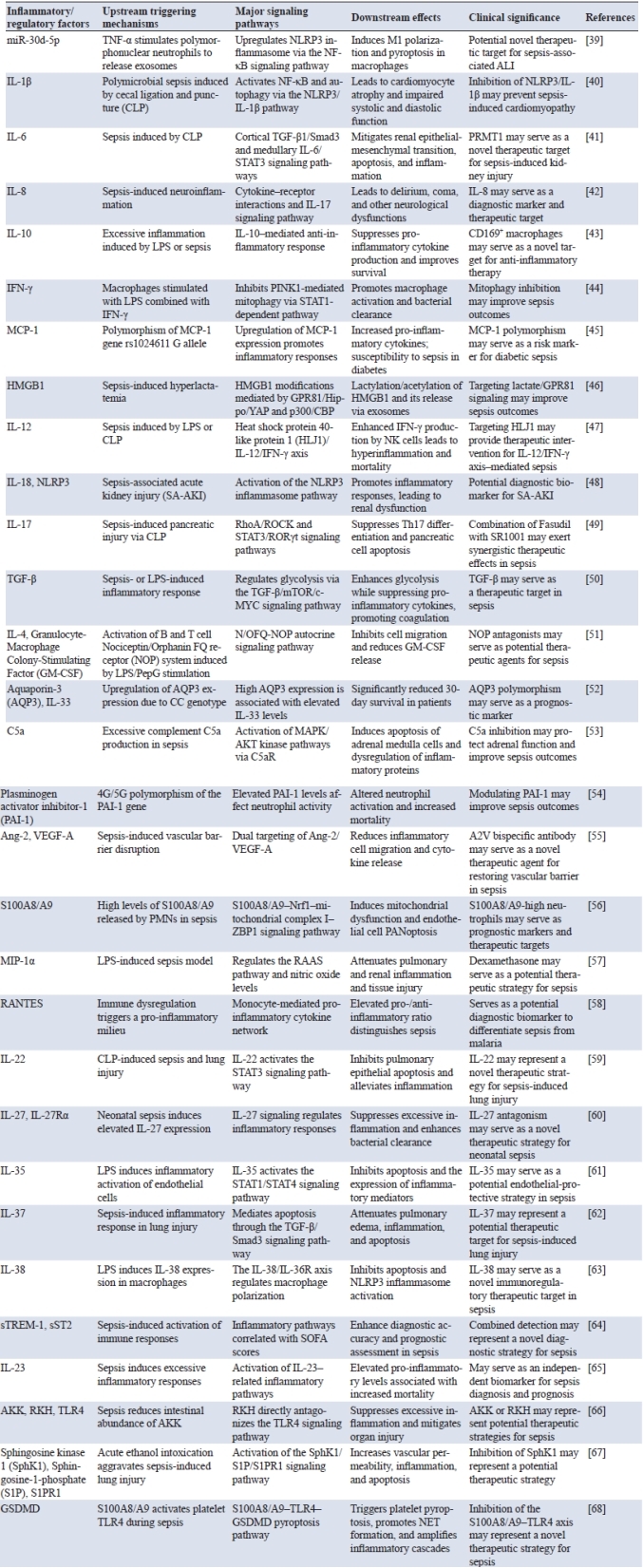
2.3 Bacterial toxins and host cell damage
Bacterial toxins and host response dysregulation induce cellular injury and organ failure. Several signaling pathways and organelle injury processes have been implicated in sepsis, indicating that they are not only part of the pathological mechanism but also potential therapeutic targets.
In skeletal muscle, SPSB1 worsens sepsis-induced muscle atrophy and regeneration failure through inactivation of the TβRI–Akt–myogenin signaling pathway and suppression of protein synthesis [69]. Intestinal ACE2 can modulate the gut microbiota and the metabolite 5-MTP, activate the PI3K–AKT–WEE1 pathway, and support barrier function, thereby mitigating sepsis and multi-organ damage [70]. In the myocardium, actin/myosin degradation by calpain-1 plays a major role in sepsis-induced heart injury. Interventions targeting calcium signaling can mitigate cardiac dysfunction and increase the survival rate [71]. Extracellular histones are also detrimental to the heart, as they cause imbalances in calcium homeostasis and mitochondrial function, resulting in cardiac failure. Inhibition of histones is a novel therapeutic strategy [72].
The impairment of both endothelial and barrier function of vasculature is especially important in the development of sepsis. Sepsis-induced disseminated intravascular coagulation disrupts EXT1/FGFR1-mediated glycocalyx remodeling and enhances barrier damage and pulmonary edema; TGF-β1 derived from platelets induces neutrophil recruitment and promotes extracellular trap formation, leading to thrombosis as well as organ injury [73]. Its absence increases survival, and makes it a candidate therapeutic target [74]. At a later stage after shock, the Angiopoietin-2 (Ang-2)/Tie2 pathway is involved in vascular leakage, and subsequently contributes to remote, indirect Acute Respiratory Distress Syndrome (ARDS).
Inhibition of Ang-2 could restore barrier function and increase survival rate [75]. Additional studies demonstrate that Ang-2 also induces lung injury and increases inflammation by modulating endothelial-neutrophil interactions [76].
Mitochondrial injury and dysfunction are key elements of sepsis-induced organ failure. Mitochondria-related hub genes have been shown to influence energy metabolism and immune microenvironment regulation, which are significantly involved in the pathogenesis and progression of Sepsis-ACM [77]. Histone Deacetylase (HDAC) 3 inhibits mitochondrial quality control by the FOXO1–Rho-associated coiled-coil containing protein kinase (ROCK) 1 axis, driving progression of acute lung injury (ALI); thus, HDAC3 inhibition might represent a promising novel therapeutic opportunity [78]. The mechanosensitive ion channel Piezo1 is triggered during sepsis to induce calcium influx and mitochondrial impairment which in turn disrupts the tight junctions of the intestinal epithelial cells, followed by cellular apoptosis, thereby propagating barrier damage [79].
Immune and reparative functions are also suppressed in the long term. Sepsis may lead to the dysfunction of bone marrow (BM) mesenchymal stem cells (MSCs), which is characterized by mitochondrial depolarization and decreased cytokine expression, thereby disrupting BM homeostasis and long-term immune reconstitution [80]. Recurrent sepsis worsens CD4⁺ T cell exhaustion, interferes with antiviral immunity, and is consequently associated with long-term mortality, demonstrating the sustained immune consequences of sepsis [81]. Furthermore, artesunate and metabolism-related proteins also show therapeutic potential by regulating hepatic endothelial cell subpopulation proportions, inhibiting inflammatory cytokine release in liver macrophages, and decreasing lymphocyte apoptosis, thereby alleviating liver injury and immune imbalance and supporting their potential clinical application [82]. VDBP is reduced with sepsis and correlated with liver injury and mortality. Its overexpression could protect against oxidative stress and hepatocyte injury, which may have a protective role [83].
Interactions between bacteria-derived molecules and host response mediators constitute central driving forces in the pathogenesis of sepsis. Emerging evidence highlights the critical role of inflammatory cytokine networks in this process. For example, the combined action of tumor necrosis factor with IL-18, IFN-γ, or IL-1β is sufficient to reproduce sepsis-associated tissue injury, indicating that host inflammatory mediators alone can drive systemic pathological alterations [84]. Concurrently, metabolic disturbances in skeletal and cardiac muscle are closely associated with endothelial barrier disruption and immune dysfunction, thereby facilitating the transition from localized inflammatory injury to systemic pathology. Sepsis is therefore characterized by a complex and highly lethal pattern of multi-organ damage, with key pathological hubs including mitochondrial dysfunction, vascular barrier breakdown, and immune exhaustion. These pathological nodes not only reveal the underlying logic of disease progression but also represent potential targets for precision therapeutic interventions. For instance, recombinant angiopoietin-like protein 4 has been shown to significantly reduce vascular leakage, organ failure, and mortality in murine models of lethal sepsis and Neisseria meningitidis infection, further underscoring the critical role of endothelial barrier integrity in disease progression [85]. When the immune system is rapidly mobilized to combat infection, this protective response can escalate into a systemic storm of signaling mediators. Inflammatory cell death and cytokine release mutually reinforce one another, forming a self-sustaining inflammatory loop (Figure 1) that drives the progression of inflammation from local dysregulation to systemic inflammatory imbalance. Within this cascade, aberrant activation of immune cells together with uncontrolled release of inflammatory mediators becomes a central force sustaining the inflammatory response, thereby laying the mechanistic foundation for subsequent discussions on immune regulation and inflammatory signaling.
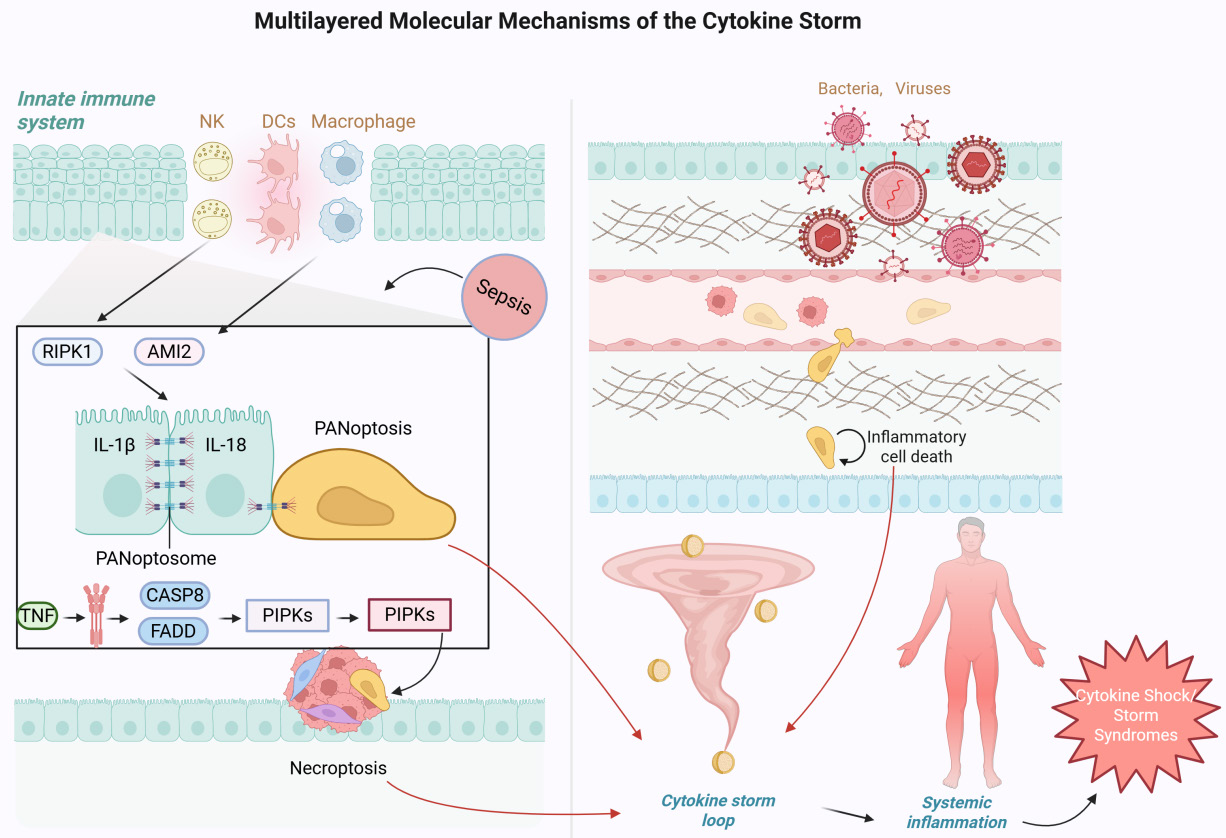
Figure 1. Multi-layered molecular mechanism of inflammatory storm. After pathogen invasion, the innate immune system is quickly activated by NK cells, dendritic cells and macrophages, inducing a vast amount of inflammatory mediators. This results in an inflammatory cascade focused on TNF, IL-1β and IL-18. The AIM2-dependent pathway works together with RIPK1 to promote the formation of the PANoptosis complex, including the assembly of PANoptosome components such as CASP8 and FADD, thus leading to inflammatory cell death and cytokine amplification. A variety of programmed cell death modalities, including pyroptosis and necroptosis, are interwoven to synergistically amplify tissue damage, as illustrated in the left panel of the figure, leading to the formation of the “cytokine storm loop”. The right panel further depicts how inflammatory cell death and cytokine release disrupt tissue barriers and propagate inflammation systemically. Such a self-perpetuating inflammatory network can transgress tissue boundaries, producing a systemic inflammatory response and ultimately contributing to multiple organ failure and cytokine shock syndromes.Created by the authors using BioRender.
3.1 Activation of immune cells and release of inflammatory cytokines
In sepsis-induced ALI and multiple organ dysfunction syndrome, the devastating cascade is mainly due to the abnormal activation of immune cells and the dysregulated release of inflammatory mediators, which triggers a harmful inflammatory cascade. Neutrophil-derived exosome miR-30d-5p was found to promote macrophage M1 polarization and pyroptosis, thus representing a critical element that drives lung injury [39].
eCIRP facilitates the generation of aging neutrophils with augmented antigen presentation, leading to Th1 differentiation and IFN-γ–dependent accelerated high-level Neutrophil Extracellular Trap formation (NETosis). This amplifies the inflammation and damages tissues [86]. Furthermore, manganese ions also enhance lipopolysaccharide (LPS)-induced innate immune responses through activation of the cyclic GMP-AMP synthase–stimulator of interferon genes pathway, which leads to severe septic shock [87]. In the nervous system, septic plasma exosomes can induce microglial activation via TLR7/MyD88 signaling and result in neuroinflammation, which is associated with the pathogenesis of sepsis-associated encephalopathy (SAE) [88]. In addition to pro-inflammatory responses, protective physiological countermeasures are present in the body. S-phase kinase-associated protein 2 (Skp2) relieves tissue injury by suppressing ferroptosis in lung epithelial cells; deletion of G protein-coupled receptor 174 (GPR174) enhances regulatory T cell (Treg) function and M2 macrophage polarization, thereby inhibiting excessive inflammation [89, 90]. Furthermore, DPP4 and TXN have been suggested as probable indicators for immune-regulation functions, which can offer new molecular targets for precision therapy [91].
The intersection of metabolism and inflammation is also important. Tripterygium wilfordii glycosides inhibit the Warburg effect via PKM2/high mobility group box 1 (HMGB1), which attenuates inflammation and organ damage, while neutrophil extracellular traps (NETs) promote ferroptosis by Methyltransferase-Like (METTL)3-mediated N6-methy-ladenosine (m6A) modification of Glutathione Peroxidase 4 (GPX4), aggravating lung injury [92, 93]. In addition, interactions among inflammatory factors are also important. TNF-α and IFN-γ induce PANoptosis through the JAK/STAT1/IRF1 axis to cause severe cell death and tissue damage. This process is also observed in COVID-19 and sepsis [94]. On the other hand, the lactate-enhanced qSOFA score has shown good predictive value for in-hospital mortality in patients with sepsis; mechanistically, LPS/IFN-γ inhibit PTEN-induced kinase 1 (PINK1)-mediated mitophagy through the STAT1-caspase pathway, thereby promoting mitochondrial reactive oxygen species production, bacterial clearance, and improved survival during sepsis [13, 44].
Immune activation in sepsis occurs through multiple layers of pro-inflammatory amplification, including extracellular exosomal miRNAs, NET formation, and PANoptosis, which collectively drive acute organ injury. However, persistent inflammatory stress progressively reprograms immune regulatory networks, shifting the host response from hyperinflammation toward immune exhaustion and dysregulation. This transition ultimately leads to the immune imbalance that defines the later stages of sepsis.
3.2 Immune system imbalance and hyperreaction
Following this storm of inflammation, there is a rapid transition toward an immunosuppressive state in sepsis patients, with the establishment of a classic “immune imbalance”. Reduced expression of HLA-DR on monocytes is a hallmark of this stage and is strongly related to poor prognosis [95]. In terms of molecular mechanisms, p53 stimulates immunosuppression through inhibition of CD4⁺ T cell proliferation and upregulation of apoptotic pathways, ultimately disrupting T cell homeostasis; the absence of Sulf-1 alters endothelial heparan sulfate modifications, resulting in glycocalyx dysfunction that mediates pulmonary immunosuppression [96, 97]. The emerging role of non-coding RNAs in regulation of immune response has been recently highlighted. lncRNA Metastasis-Associated Lung Adenocarcinoma Transcript 1 (MALAT1) modulates polymorphonuclear myeloid-derived suppressor cell (PMN-MDSC) differentiation via STAT3 phosphorylation, and its dysregulation enhances immunosuppressive functions [98]. Meanwhile, MLIP alleviates excessive inflammatory responses by regulating macrophage metabolism and oxidative stress, providing a potential target for restoring immune balance [99]. In dendritic cells, SREBF1 mediates lipid metabolism reprogramming and endoplasmic reticulum stress, thereby driving immune paralysis and further locking the body into an immunosuppressive state [100].
The diversity of immunotypes has also been confirmed by population-based studies. Transcriptomic analysis divides sepsis into two subgroups, SRS1 (immune-suppressive type) and SRS2 (predominantly proinflammatory type). SRS1 is classically defined by low HLA-II and T cell exhaustion, and is strongly associated with lower survival rates [7]. Persistent upregulation of immune checkpoint molecules, including PD-L1–related inhibitory pathways, contributes to T cell exhaustion and impaired antiviral responses, reinforcing the immunosuppressive phenotype in sepsis [81, 101, 102].
Therapies targeting immune exhaustion have shown promise. Nicotinamide riboside supplementation has been shown to partially restore T cell function via activation of NAD⁺/SIRT1 signaling pathways, restore immune response, and decrease organ injury and mortality [103]. However, in elderly patients, prolonged elevation of IL-6 and increased T cell exhaustion contribute to a more sustained immunosuppressive state that is less amenable to intervention [104]. Notably, ILC2-derived IL-13 secretion in response to PD-1 deficiency partially reversed long-term muscle weakness and increased gene expression in the skeletal muscle. This indicates a strong relationship between immunosuppression and long-term functional deficits [105].
In the escalating inflammatory cascade, the immune response shifts toward a biphasic dysregulated state characterized by concurrent hyperactivation and immunosuppression. Crosstalk between pro-inflammatory and immunosuppressive signals shapes a complex immune landscape (Figure 2), driving sepsis progression and the transition of host defense from coordinated activation to dysregulated exhaustion. Within this network, diverse inflammatory mediators regulate intercellular communication, immune activation, and feedback control. For example, the monocyte-associated molecules CAP-1 and IL-16 participate in cell–cell communication in sepsis and septic shock and have been proposed as potential diagnostic biomarkers [106]. Elucidating the roles of inflammatory mediators in this regulatory network may therefore provide insights into sepsis pathophysiology and therapeutic targeting.
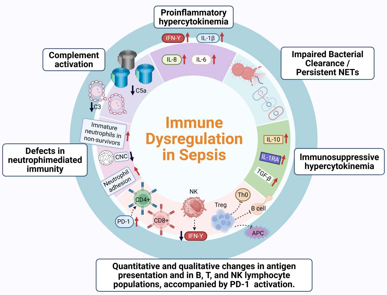
Figure 2. Pathophysiological mechanisms of immune imbalance in sepsis. The immune dysregulation induced by sepsis is a biphasic response in which proinflammatory and immunosuppressive phases succeed each other, as illustrated in the circular schematic showing interconnected immune pathways. At the very beginning, over-activation of the complement system (C3, C5a) and neutrophil dysfunction amplify inflammation to an overwhelming level, as exemplified by a dramatic rise in factors like IL-1β, IL-6 and IFN-γ, corresponding to the upper proinflammatory hypercytokinemia segment. Then,
immunosuppressive cytokines (IL-10, IL-1RA, TGF-β) release follows, leading the host into a condition of immune exhaustion and chronic infection, as depicted in the right immunosuppressive hypercytokinemia segment, which is also associated with impaired bacterial clearance and persistent NETs. Alterations of B, T and NK lymphocytes in terms of their numbers and quality, as well as uncontrolled activation of the PD-1 pathway, together represent patterns most likely to lead to failed antigen presentation and subsequent immune paralysis, as shown in the lower portion of the figure highlighting lymphocyte dysfunction. This depicts a time-dependent progression of systemic inflammation to immunosuppression that forms the unmodifiable pathological basis for sepsis. Created by the authors using BioRender.
3.3 The role of inflammatory mediators in sepsis
Inflammatory mediators in sepsis function as a double-edged regulatory network that amplifies immunity while inducing negative regulation. Pyroptosis-related extracellular vesicles (pyroEVs) carrying membrane-expressed ASC proteins attach to B cells and suppress TLR4 signaling, reducing overactivated inflammation. This indicates that vesicle-mediated interventions might be of potential therapeutic value [107]. In contrast, ferritin promotes NETosis in an MSR receptor-dependent manner via the Peptidylarginine Deiminase 4 (PAD4)/NE/ROS pathway, resulting in aggravated cytokine storm and lung injury, marking its important pro-inflammatory role in the process [108].
Among inflammatory cytokines, higher levels of MIP1α and IL-1ra are associated with lower mortality, suggesting that some mediators may counterbalance dysregulated inflammatory responses and serve as potential therapeutic targets [109]. Dexmedetomidine exerts protective effects on neuroinflammation and blood-brain barrier disruption by promoting astrocyte α2A receptor activation, implying the dynamic homeostasis of inflammatory regulation circuits between inflammation and the nervous system [110]. The enrichment of CD14⁺CD16⁺ monocytes in the blood, on the other hand, correlates tightly with increased susceptibility, severity, and mortality in sepsis, highlighting an essential role for disequilibrium among cellular populations in immunopathology [111].
Both genetic and clinical investigations support the predictive and stratification value of inflammatory variables. TNF-α -308 AA genotype or the carriers of the A allele is associated with higher serum TNF-α levels, and may serve as a genetic predictor of inflammatory responses [112]. Plasma TNF-α levels in septic patients are about 10 times higher than those in healthy individuals [113]. The TNF-α -308G/A polymorphism and IL-6 levels do not correlate with mortality in neonatal sepsis, although the prediction capacity may be heterogeneous in the population and age [114]. Of interest, TNF-α level was significantly higher in children with positive blood cultures, particularly those with Gram-negative bacterial infection and could be another alternative marker for early intervention [115]. Moreover, elevated levels of IL-6 and IL-8, as well as of KC-like and Regulated upon Activation, Normal T Cell Expressed and Secreted (RANTES), have been observed in experimental models, demonstrating the conservation of inflammatory mediators during immunopathology and their application to clinical research [116].
Inflammatory mediators in sepsis not only drive the cytokine storm and organ injury but also provide important clues for risk prediction, patient stratification, and precision therapies. The production and immunoregulatory functions of these mediators rely on the activation of multiple cellular signaling pathways, which play critical roles in inflammatory amplification, immune imbalance, and organ damage. Within this complex signaling network, N-myc and STAT interactor (NMI) has been identified as an important regulator that integrates inflammation-related signals from multiple receptors. Studies have shown that NMI participates in immune signaling through receptors such as IFNAR1, TNFR1, and TLR family members, and is closely associated with the activation of NF-κB, MAPK, and JAK pathways, thereby promoting pro-inflammatory cytokine expression and enhancing inflammatory responses [117]. Among these pathways, Toll-like receptor (TLR) signaling represents a central mechanism linking pathogen recognition to downstream inflammatory cascades in sepsis.
4.1 TLRs and signaling cascades
As the central sensors of the innate immune system, TLRs are essential in promoting sepsis-associated inflammatory storm and immune dysregulation. Their signaling pathways are critical not only for pathogen recognition but also for inflammasome activation, metabolic imbalance, and organ failure during sepsis. There is abundant experimental and clinical data demonstrating that TLR signaling pathways play a dual role in amplifying inflammation and mediating therapeutic intervention. Fh15 protein derived from a parasite and small molecule inhibitor TAK-242 effectively inhibit LPS-induced inflammation and tissue injury, which indicates that TLR4 can be a therapeutic target [118, 119]. In parallel, both bacterial mRNA and LPS work in concert to activate the NLRP3 inflammasome, with NLRP6 also undermining host defense by sensitizing T cells through the IL-18 release. This implies a complicated interrelation and potentiation of TLR signaling by inflammasomes [120, 121].
At the systemic level, the MyD88–NF-κB–TNF-α pathway manifests a rapid phase-specific activation pattern in response to polymicrobial infection while malignant fibrous histiocytoma amplified sequence 1 presented the features of early inhibition combined with late progression along the TLR2 pathway. Such observations allow for a temporal view of the course of sepsis [122, 123]. Furthermore, serum amyloid A (SAA) acts via TLR2/TLR4 to transform HDL into pro-inflammatory particles, enhancing vascular inflammation. The elevated expression of genes like LCN2 and ELANE also support a role of TLR pathways in the immune dysregulation [124, 125]. Of note, the contribution of TLR signaling goes beyond inflammation and affects metabolism and endocrinology. TLR4/MD2-mediated LPS suppresses the growth hormone receptor, demonstrating its distinctive role in metabolic adaptation during sepsis [126]. In contrast, the transcription factor ZBTB20 enhances tissue damage through augmented activation of NF-κB. Its depletion greatly ameliorates inflammatory injury, thus emerging as a novel therapeutic target [127].
TLR-associated signaling pathways are not only central initiators of the inflammatory storm in sepsis but also important links between immunosuppression and metabolic disorders. A comprehensive exploration of their dynamic properties and their interplay mechanisms may establish the basis for the discovery of the intact landscape of disease development, as well as novel strategic avenues for stratified management and precision intervention in sepsis.
4.2 Cytokine storm and signal amplification
Excessive cytokine production represents a hallmark of immune dysregulation in sepsis, and failure to contain signal amplification contributes to systemic inflammation and multiple organ failure. Research has indicated that TNF-α and IFN-γ may cooperatively facilitate PANoptosis depending on the JAK/STAT1/IRF1 axis, which can be responsible for the deadly inflammatory cascade in COVID-19 and sepsis [94]. The systemic inflammatory response syndrome in fetal sepsis is responsible for multiple organ dysfunctions and is directly linked to severe neonatal morbidity, sepsis-related mortality and long-term sequelae risk [128]. It has also been shown that the combined action of pro-inflammatory cytokines like TNF and IL-18 can mirror many systemic effects of sepsis, leading to pervasive rewiring in both gene transcription and cellular composition that amplifies tissue destruction [84].
In the past few years, the targeting/inhibition of inflammation-induced cell death has been proposed as one of the major advances in controlling cytokine storms. Rapamycin has been reported to alleviate endothelial injury by promoting autophagy while suppressing gasdermin D (GSDMD)/caspase-1-mediated pyroptosis and decreasing the production of pro-inflammatory cytokines IL-1β, IL-18 [129]. The caspase-1 inhibitor AC-YVAD-CMK can also ameliorate acute kidney injury (AKI) by inhibiting the NLRP1/GSDMD pathway [130]. In contrast, the MSR-dependent PAD4/NE/ROS pathway in macrophages triggers NET formation in response to high levels of ferritin and promotes inflammation and lung injury, representing a mediator of poor prognostic significance [131].
The immunological features of cytokine storms vary across diseases. Sepsis and hemophagocytic lymphohistiocytosis (HLH) are diseases of overwhelming inflammation, but in contrast to sepsis patients, HLH patients show significant overactivation of CD38high/HLA-DR+ effector T cells, primarily CD8+ T cells. Conversely, this phenotype is not seen in early sepsis, providing a molecular rationale for the clinical distinction between the two entities [132]. Moreover, the Epidermal Growth Factor Receptor can transactivate and induce production of TNF-α in cardiomyocytes through the TACE/TGF-α pathway. Its inhibitor has been demonstrated to potently reverse heart dysfunction in animal subjects and supports the existence of druggable signaling nodes in the myocarditis inflammation network [133].
Additional layers of regulatory axes are also being uncovered. The hepatocyte Pannexin 1 (PANX1)–IL-33 axis is triggered by the ATP–P2X7 pathway, leading to ST2+ Treg infiltration. This axis acts as a negative regulator for inflammation amplification, attenuating endotoxin-induced liver injury [134]. Resveratrol inhibits the microglial NLRP3/IL-1β axis via receptor-dependent mechanisms, improving cognitive function in SAE; phosphodiesterase 10A inhibitors suppress pyroptosis and organ damage by inhibiting NLRP3 inflammasome activation, providing a new idea for neuroprotection [135, 136]. Furthermore, the natural product EGCG may ameliorate inflammation and ferroptosis by dampening RELA activity and suppressing exaggerated macrophage activation, indicating that plant-derived molecules may have therapeutic potential for inflammation-related intervention [137]. IL-6 signaling, as another core pathway, regulates microtubule dynamics through the JAK2/Ninein axis, thereby promoting Extracellular Signal-Regulated Kinase (ERK) 1/2-mediated phosphorylation of p47phox and LC3-associated phagocytosis. Its deficiency leads to immune paralysis and dysfunction of monocytes/macrophages [138]. This finding not only demonstrates the dynamic changes of inflammation amplification and immune hyporesponsiveness, but also offers a novel mechanistic basis for precision intervention.
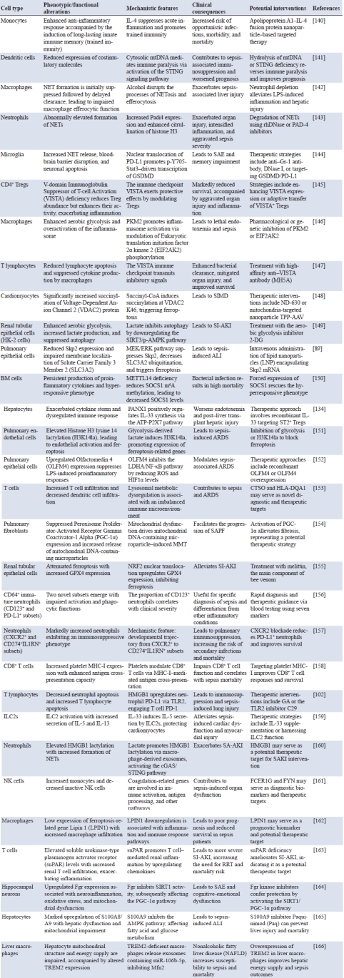
Cytokine storms drive uncontrolled inflammation and multi-organ injury in sepsis, and their cross-pathway amplification reflects the dynamic imbalance between excessive immune activation and compensatory immunosuppression. Computational modeling that captures key signaling nodes and their spatiotemporal dynamics has emerged as a valuable framework for understanding these complex inflammatory networks and identifying potential therapeutic targets. In particular, agent-based modeling (ABM) has been applied to simulate systemic immune responses and to explore fundamental mechanisms of sepsis pathogenesis [139]. Persistent immune dysregulation is accompanied by profound phenotypic and functional remodeling of immune cells, including alterations in metabolic programs, differentiation trajectories, and signaling regulation, collectively shaping the systemic landscape of immune exhaustion (Table 2). Table 2 summarizes the major functional switches and signaling perturbations of key immune cell populations during sepsis, providing a cellular framework for understanding immune imbalance and setting the stage for further discussion of immune cell–specific regulatory mechanisms.
4.3 Negative feedback mechanisms and immune homeostasis
In addition to the systemic uncontrolled inflammatory response, sepsis is also characterized by the activation of various negative feedback mechanisms that serve to curb tissue damage and maintain immune homeostasis. Inhibition of NLRP3 inflammasome activation and pyroptosis by certain metabolic products of myeloid cells, including itaconate, promotes inflammatory tolerance and limits tissue damage [167]. Serum exosomes have a dynamic molecular pattern in the progression of disease and their correlation with inflammatory, coagulation and vitamin metabolism pathways makes them a new approach to integrate diagnosis and therapy [168].
Perturbations in immune homeostasis are associated with disease progression. Silencing Cystatin C accelerates LPS-induced inflammation by inhibiting autophagy and increasing caspase-11 expression, which enhances NLRP3 inflammasome activation [169]. T-cell lymphoma breakpoint-associated target genes and Sestrin 2 boost host resistance and disease tolerance by inducing autophagy and mitochondrial quality control, which increases clearance of bacteria but attenuates organ damage [136, 170]. These data demonstrate that autophagy-associated pathways are central hubs for fine-tuning inflammation versus tissue protection.
In terms of signaling regulation, S100A10 inhibits the TLR pathway to downregulate excessive inflammation, and lack of it increases immune damage [171]. Membrane-bound MHC I molecules feed back to suppress TLR signaling through the Fps–SHP-2 pathway to provide self-restricted protection of innate immunity [172]. Meanwhile, immunosuppression may stem from imbalanced endogenous negative feedback, whereby SAMSN1 promotes an immune-suppressive state that can be reversed upon its depletion [173]. APE1 deficiency impairs macrophage phagocytic ability via the GSK3β/Nrf2 axis, contributing to bacterial infection and immune defects [174]. Notably, TSLP signaling serves as a central protective factor for myeloid cells. Its absence does not impair bacterial eradication, but exacerbates systemic inflammation and organ injury. By contrast, the survival of mice is significantly enhanced by TSLP-mediated suppression of hyperactivation of myeloid cells [8].
While dysregulated signaling cascades orchestrate the acute immune and metabolic responses in sepsis, inter-individual variability in these pathways is profoundly influenced by inherited genetic variations. Genome-wide association studies have identified several low-frequency variants associated with reduced 28-day survival in sepsis, including a missense variant in SAMD9 (hazard ratio [95% CI]=1.64 [1.37–6.78], P=4.92×10⁻¹⁰), a gene implicated in inflammatory responses that may mediate tissue injury [175]. In parallel, integrative genetic analyses incorporating Mendelian randomization have identified multiple candidate genes significantly associated with sepsis susceptibility, several of which remained robust after sensitivity analyses. These findings collectively highlight the contribution of host genetic variation to disease susceptibility and prognosis [176]. Thus, integrating host genetic architecture into the framework of signaling networks provides an essential dimension for understanding disease heterogeneity and differential clinical outcomes. Importantly, many of these genetically influenced pathways also generate measurable molecular signatures that are increasingly investigated as emerging biomarkers for diagnosis, risk stratification, and prognosis in sepsis.
5.1 Genetic polymorphisms and individual differences
Host genetic variations play a critical role in sepsis susceptibility and outcomes, affecting various aspects ranging from pathogen recognition and immune responses to metabolic homeostasis and organ injury. Matrix metalloproteinase (MMP) 8 rs11225395 G/G genotype is associated with sepsis susceptibility, and MMP1 and MMP3 are not only linked to bacterial and viral infection susceptibility but also to agent-specific levels of severity. This indicates that the genetic variation of the host plays a role in pathogen-specific patterns of immune defense [177]. Gene-based analyses have also demonstrated that sepsis-2 and the Sepsis-3 criteria not only have a polygenic overlap but also possess unique molecular attributes. Their multigene risk scores also have a high predictive value, suggesting that sepsis susceptibility usually results from the combined effect of multiple small-effect polymorphisms [178].
The interplay of genetic susceptibility and environmental factors has been given more attention in research. For instance, genetic insomnia is positively associated with sepsis risk, and this link is in part explained by cardiac metabolism pathways involving heart function. This indicates that sleep disturbances may be a targetable genetic risk factor [179]. Alterations of the coagulation and complement cascades contribute significantly to the disease course. Polymorphisms in the THBD gene are strongly associated with AKI risk and death [180]. A gain-of-function variant in cholesteryl ester transfer protein (CETP) is associated with increased acute sepsis mortality, whereas genetic evidence for decreased CETP function, together with pharmacological CETP inhibition in humanized mouse models, suggests potential survival benefits through the preservation of HDL-C levels [181]. Different variants of complement C5 can either reduce susceptibility or, conversely, accelerate disease progression by enhancing C5a generation and the release of pro-inflammatory mediators [182].
Immunity- and metabolism-related pathways also serve as hub nodes that regulate the disease. The VDR Fok I polymorphism is a strong risk factor for sepsis, and it correlates positively with lower vitamin D status, thus indicating that the vitamin D metabolic axis may represent an important host regulatory pathway [183]. In patients with diabetes, an association was found between Monocyte Chemoattractant Protein-1 (MCP-1) rs1024611 G allele and susceptibility to sepsis, which may be attributed to upregulated expression of MCP-1 and TNF-α [45]. High-risk allele combinations of TNFA rs1800629 and TLR4 rs4986791 in trauma patients are associated with significantly increased risk of sepsis, considering that TLR4 is a prognostic variant [184]. Moreover, although the reported associations of certain polymorphisms in genes including CHRNA7, NR5A2 and SUFU with sepsis-associated acute kidney injury (SA-AKI) have not shown genome-wide significance, these findings underscore the influence of host genetic differences on disease phenotype [185]. Polymorphisms in inflammation-related genes provide broader mechanistic insights. The elevated serum and urinary IL-10 levels in carriers of the IL-10 rs1518111 T allele are strongly associated with the development of SA-AKI, demonstrating that both inflammatory factor levels as well as genetic susceptibility play critical roles in the risk of organ damage [186]. Furthermore, PCSK9 loss-of-function not only increases organ injury by altering lipoprotein metabolism and removal of endotoxins but also impairs endothelial function by regulating Angpt-1, thereby aggravating septic pathology [187].
Susceptibility to sepsis is not monogenic but due to the combined action of multiple pathway variants. Such effects include inflammation control, immunological response, coagulation deregulation and metabolic perturbation. A comprehensive investigation of the dynamic properties and interaction mechanisms in these genetic networks can not only help uncover the molecular pathogenic basis for sepsis susceptibility, but also serves as an essential framework for developing risk prediction models and precision intervention strategies.
5.2 Gene expression regulation and inflammatory response
Inflammatory dysregulation in sepsis does not ensue from a single signal but rather represents a systemic disorder arising as a consequence of the perturbation of transcriptional homeostasis, abnormal epigenetic changes and malfunctioning noncoding RNA networks. It has been reported that the disproportion of expression of TLR4 and GRP78 is closely related to patient prognosis, indicating their dynamic properties during abnormal inflammatory responses and their potential to serve as monitoring parameters [188]. At a much deeper level, however, the transcription factor PU.1 induces MDSC differentiation and aggravates immune suppression via lncRNA Hotairm1 and its related epigenetic modification network. This connects transcription factors, epigenetic marks and immune dysregulation in a coordinated manner [189]. Additionally, the function of m6A methylation in immune cell infiltration and Th17 differentiation is receiving more attention. Master factors, such as METTL16 and IGF2BP1, were demonstrated to be involved in the progression of the late inflammatory phase, strongly suggesting the utility of epigenetic manipulation [190].
Multivariate complexity of gene regulatory networks in organ injury varies across different organs. Adenosine-uridine RNA editing of neuronal gene Grik2 and Flna in SAE are involved in the development of cognitive deficits. This phenomenon exhibits age- and sex-specific traits [191]. Research on natural compounds has discovered new ways of modulating inflammation and oxidative stress.Vitexin relieves lung injury in part through the SNHG1/DNA methyltransferase (DNMT) 1/miR-495 axis, while Gossypol alleviates myocardial injury via HDAC inhibition and histone acetylation remodeling [192, 193]. Furthermore, exosomes and non-coding RNAs were found to be implicated in immune reprogramming. Microvesicles derived from patients can deliver DNMT1/3A, which results in hypermethylation of the TNF-α promoter and immunosuppression [194]. circMAPK1 promotes NLRP3 inflammasome activation through modulating demethylation, which worsens lung injury [195]. On the other hand, miR-221-5p enhances inflammatory responses by inhibiting mitophagy and promoting organ damage as well [196].
The widespread involvement of m6A modification in sepsis-induced multiple-organ injury is noteworthy. METTL3-triggered methylation has been reported to promote liver injury progression, but in the heart, lncRNA and mRNA expression differences lead to functional decline through the TNF and PI3K-Akt signaling pathways [197, 198]. In AKI, the NF-κB/miR-376b/NFKBIZ negative feedback system not only controls inflammation and cell death but also shows the diagnostic significance of urinary miR-376b [199].
Gene regulatory networks of sepsis have global characteristics with complex coupling between multiple hierarchical levels, pathways, and organs. In this dynamic, feedback-loop-driven environment, inflammation potentiation and immune dysfunction maintain a parallel course, leading to multi-organ failure. More detailed exploration of these mechanisms not only uncovers the molecular basis of sepsis but also provides a basis for precision therapy and personalized medicine.
5.3 Clinical significance of emerging biomarkers
Advances in novel biomarkers are reshaping the diagnosis and treatment of sepsis. Evidence from the endocrine and metabolic aspects suggests that using T3 within the 60–80 ng/dl range is related to lower incidence rates of Persistent Inflammation, Immunosuppression, and Catabolism Syndrome (PICS), whereas profound T3 deficiency in combination with high Sequential Organ Failure Assessment (SOFA) scores predicts poor outcomes [200]. Metabolomic studies have indicated the potential causal role of dysregulation of the glycolysis/pyruvate pathway in the risk and prognosis of sepsis, which can lay a strong foundation for developing metabolic biomarkers for early diagnosis [201].
Genetic and immune biomarkers also show significant clinical utility. In patients, individuals with the LAG-3 rs951818 AA genotype were found to have a reduced risk of death, suggesting their potential value as a prognostic genetic marker [202]. Furthermore, molecular fingerprints of IRRGs perform well in identifying high-risk individuals and monitoring immune status, paving the way for precise intervention [203]. Synergistic detection of inflammatory mediators has also improved clinical prediction models. IL-10 levels integrated with the National Early Warning Score (NEWS) score further enhances sensitivity in early detection of sepsis risk, and the combined measurement of urinary CRP, α1-acid glycoprotein and SAA dramatically improves discrimination between sepsis and SIRS [204, 205]. In addition, easy-to-obtain markers such as NLR, PLR and LMR have been shown to be clinically useful in the early postoperative period [206]. Furthermore, the T-cell CD69 ratio combined with PCT can accurately differentiate between gram-negative and gram-positive sepsis and also has predictive value [207].
The range of biomarkers is constantly increasing based also on multi-omics and non-coding RNA research. Excessive expression of CEACAM8, MPO and RETN has been demonstrated to be closely correlated with disease progression, hence offering potential as diagnostic urinary biomarkers [208]. In addition, the correlation between PGK1 and immune cell infiltration and inflammation regulation also demonstrates its potential as a new diagnostic index [209]. Furthermore, the roles of ncRNAs have become a subject of much interest. LINC01278 serves as a sponge of miR-451a to weaken inflammation and organ injury, and MEG3 rs7158663 GG genotype dramatically elevates genetic susceptibility and adverse outcome risk through promoting the overflow of inflammatory factors [210, 211]. Together, these findings depict a multidimensional biomarker network encompassing metabolic, genetic, immune, and non-coding RNA pathways. This network not only deepens our understanding of the complex molecular landscape of sepsis but also facilitates improved patient stratification and immune status monitoring. Importantly, these advances provide a critical basis for the development of precision medicine approaches and targeted therapeutic strategies, paving the way for the emerging immunomodulatory and adjunctive therapies discussed in the following section.
6.1 Targeted immunomodulatory therapies
Recently, the investigation of therapeutic immune modulation has become a major trend in sepsis research. Promising approaches such as small molecules and targeted agents were discussed. For example, PLX5622 prevents neuronal injury and synapse loss by suppressing microglial activation, attenuating SAE [212]. However, the PSMA4-associated protein degrader has been identified as a new target for therapy and may reduce the risk of sepsis development [213]. Among traditional pharmacological strategies, glucocorticoids in combination with fludrocortisone have been evaluated in a component network meta-analysis of 33 randomized controlled trials including 9,898 patients, in which this combination was associated with reduced short-term mortality (RR 0.89) and improved long-term mortality in adults with sepsis and septic shock, highlighting their potential as an adjunctive therapy [214]. In addition, a prospective multicenter study of granulocyte–monocyte adsorption apheresis in 82 patients with sepsis demonstrated a significant reduction in SOFA score at 7 days and a 28-day mortality rate of 7.8%, supporting its safety and potential adjunctive benefit [215].
Moreover, the profiling of immune-cell functions has also identified additional possibilities for therapeutic intervention. Single-cell immune profiling further demonstrates the heterogeneity of immune cells in PICS post-sepsis, which offers potential individual targets for risk evaluation and precision treatment [216]. At the molecular pathway level, caspase-11/GSDMD was found to regulate NET generation via neutrophil activation, which indicates GSDMD may be a therapeutic target for sepsis treatment [217]. By contrast, HMGB1 can act as a ligand for Tim-3 to elicit immunosuppression via inhibition of NF-κB signaling in CD4+ T cells, thus representing a potential translational impact [218]. Metabolism-regulating molecules like NLRC3 mediate immunosuppression by shaping macrophage metabolism, and the targeted intervention can possibly restore the immune derangement during sepsis [219]. A CXCR2⁺ neutrophil subset has been demonstrated to promote pulmonary immune dysregulation, and its blockade could help in mitigating lung injury and improving outcomes [157]. Furthermore, ulinastatin also exerts unique immunomodulatory effects through dual regulation of neutrophils and MDSCs [220].
Platelets, T cells and metabolic pathways have also been identified as potential therapeutic targets. Platelets modulate the function of CD8⁺ T cells through MHC-I-mediated cross-presentation of antigens, and this newly described mechanism offers new opportunities in immunotherapeutic approaches [158]. IFN-γ can also restore immunosuppression by promoting a switch to the Warburg effect through the PI3K/Akt/mTOR pathway, and Inducible T-cell Co-Stimulator (ICOS)-Fc bimodulates the ICOS–ICOSL signaling axis, significantly ameliorating systemic inflammatory response syndrome and multiple organ injuries [221, 222]. Interventions on cellular and molecular levels still increase. Hematopoietic stem cell infusion, for instance, can increase MDSCs and suppress inflammation to improve survival; low-dose esmolol attenuates T cell apoptosis and normalizes Th1/Th2 imbalance by acting on the Akt/Bcl-2/Caspase-3 and ERK1/2 pathways [223, 224].
Promising microorganism and non-coding RNA-mediated therapies are also emerging. Tripeptide Arginine-Lysine-Histidine (RKH), derived from Akkermansia muciniphila (AKK), reduces inflammation and organ damage by inhibiting TLR4 signaling [66]. Nicorandil alleviates myocardial injury through mediating ferroptosis of the TLR4/SLC7A11 pathway; low-dose mycophenolate mofetil reinforces anti-bacterial activity of macrophage, hence promoting bacterial clearance for survivors [225, 226]. Molecularly, miR-150 supplementation can reverse the immunosuppressive role of MDSCs as an innovative intervention strategy for late-stage sepsis; additionally, in preclinical caecal ligation and puncture (CLP) models, umbilical cord–derived MSCs and their conditioned media were shown to mitigate organ injury, enhance bacterial clearance, reduce serum lactate and pro-inflammatory cytokine levels, and improve survival when administered within a defined therapeutic window [227, 228].
The paradigm of sepsis immunoregulation is shifting from single-target interventions toward systemic reprogramming strategies. Emerging evidence indicates that sepsis induces metabolic reprogramming and dysregulated IL-17 production in CD4⁺ T cells, leading to persistent immune dysfunction and impaired host defense after recovery [9]. These findings highlight the critical role of immunometabolic imbalance in sepsis pathophysiology and have stimulated increasing interest in therapeutic strategies targeting immune and metabolic dysregulation.
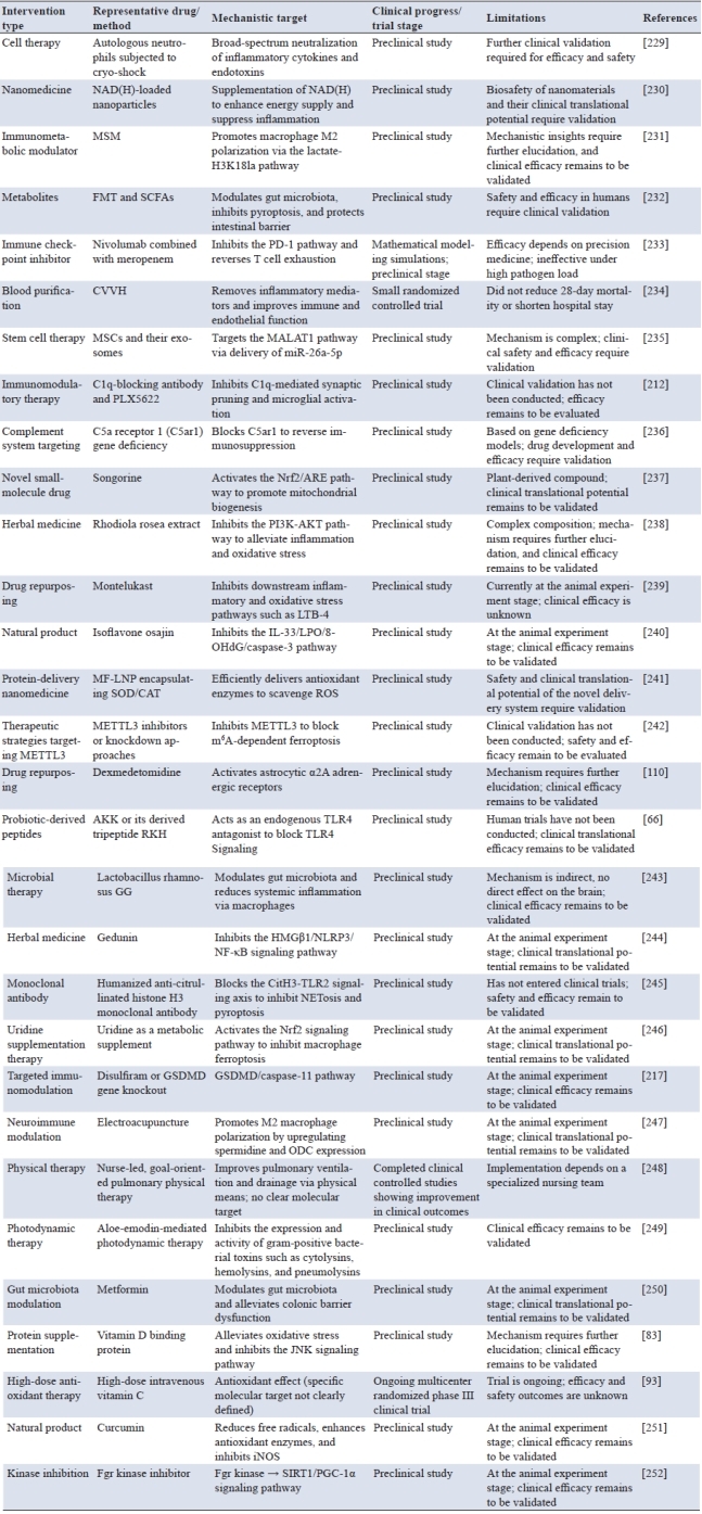
Within this evolving framework, diverse therapeutic approaches converge on a common objective: restoring immune homeostasis and cellular energy balance. Current strategies include cell-based therapies, nanodelivery systems, metabolic reprogramming, and targeted immunomodulation (see Table 3), reflecting a growing trend toward multi-target and system-level interventions. For example, mitochondrial transplantation has been shown to reverse sepsis-induced metabolic suppression and restore immune cell bioenergetics, promoting recovery of oxidative metabolism and biosynthetic capacity [253]. Beyond metabolic modulation, therapeutic strategies targeting inflammatory responses and oxidative stress are also emerging as promising approaches in sepsis management.
6.2 Novel approaches in antioxidant and anti-inflammatory therapy
In recent years, different therapeutic options for sepsis have emerged, and several promising new strategies have appeared with great clinical potential. Plasma exchange has been evaluated in a meta-analysis of five controlled trials, in which it was associated with reduced all-cause mortality in adults with severe sepsis (OR 0.54), although heterogeneity among studies was substantial and larger randomized trials are still required to confirm these findings [254]. Early enteral nutrition has emerged as a promising and generally safe therapeutic strategy in critically ill patients with sepsis and septic shock. A systematic review including five randomized controlled trials (n=442) and ten non-randomized studies (n=3,724) suggested that early enteral nutrition was associated with fewer days of mechanical ventilation and lower SOFA scores during follow-up, although no statistically significant reduction in in-hospital mortality was observed [255]. The immunomodulator glatiramer acetate (GA) has demonstrated anti-inflammatory and immunomodulatory effects in a CLP murine model of sepsis. Acute administration (1–2 mg/kg) significantly improved 72-hour survival and reduced pro-inflammatory cytokine levels, suggesting potential therapeutic relevance that warrants further clinical investigation [256].
In anti-infective therapy, the combination of azithromycin with exosomes provides a new therapeutic agent for sepsis by boosting infection clearing and regulating inflammation [257]. Nutmeg extract attenuates platelet desialylation and clearance by suppressing sepsis-elicited sialidase activity, indicating its prospect for use in the treatment of sepsis and sepsis-associated thrombocytopenia [258]. In addition, oXiris continuous hemofiltration adsorption was evaluated in a retrospective cohort of 90 patients with sepsis or septic shock. Significant improvements in hemodynamic parameters, lactate levels, inflammatory biomarkers (procalcitonin and IL-6), and SOFA scores were observed at 12 and 24 hours post-treatment. Pre-treatment SOFA score, percentage SOFA reduction, and age were identified as independent predictors of ICU mortality. Although these findings suggest potential benefits in organ function stabilization, prospective randomized trials are needed to confirm survival impact [259].
Recently, immunomodulatory therapy has become an innovative approach to the treatment of sepsis. For instance, hPF4 complexes with cfDNA and NETs to suppress thrombosis and endothelial injury, which could be potential targets for immune therapy in sepsis [260]. Codonopsis Pilosula Extract reduces immune organ damage in septic rats, and keeps the immune homeostasis by regulating BCR signaling pathway and glycerophospholipid metabolism, highlighting its future potential for sepsis immunomodulation [261]. For sepsis-related lung injury, montelukast mitigates lung tissue damage, and its administration effectively ameliorates the polymicrobial sepsis‑induced increase in inflammatory mediators and oxidative stress, thereby providing protection [239]. Gedunin exhibits a marked protective effect against oxidative stress and inflammation by suppression of HMGB1/NLRP3/NF-κB signaling pathway in septic mouse models, which provides valuable implications for its promising therapeutic applications [244]. Songorine can act through Nrf2/ARE and Nrf1 signaling pathways to enhance mitochondrial biogenesis, ameliorate cardiac function, and provide protective effects against sepsis-induced myocardial injury [237].
In the treatment of kidney injury, Osajin mitigates sepsis-induced acute kidney injury (SI-AKI) by suppressing the IL-33/Lipid Peroxidation (LPO)/8-hydroxy-2’deoxyguanosine (8-OHdG)/caspase-3 pathway and promoting antioxidant defense, indicating its possible use in sepsis therapy [240]. The deletion of MALAT1 controls epigenetic regulation, activates the methionine cycle, and improves antioxidative activity, which may provide a new direction for the treatment of sepsis [262]. Moreover, betaine remarkably decreases the levels of inflammatory and oxidative stress markers, improves gas exchange and imaging findings as well as protective effects against sepsis-induced ARDS [263].
Regarding cardiac preservation, the Mitochondria-targeted 2,2,6,6-tetramethylpiperidine-1-oxyl (MitoTEMPO) enhances mitochondrial biogenesis and protects against sepsis‑induced renal injury by decreasing mROS and IL‑1β [264]. MSCs and their exosomal miR‑26a‑5p delivery target and suppress MALAT1 to alleviate hepatocyte death and oxidative stress, and are protective against sepsis‑induced ALI [235]. Furthermore, matairesinol is a potent neuroprotectant against sepsis-induced brain damage by activating the AMPK/Nrf2 signaling pathway and blocking MAPK/NF-κB pathways to reduce both neuroinflammation and oxidative stress [265].
In terms of neuroprotection, quercetin also markedly mitigates SAE through the suppression of the CXCL2/CXCR2 axis, associated microglial activation, and neuronal ferroptosis [266]. Eriocitrin protects against SA-AKI through the activation of Nrf2 pathway, mitochondrial dynamics, and decrease in inflammation and oxidative stress [267]. Taohe Chengqi Decoction can effectively suppress the inflammatory response and correct immune imbalance in sepsis via mediation of several signaling pathways, such as MAPK and STAT3, thereby representing a potential therapeutic strategy for sepsis [268].
Emerging immunomodulatory and antioxidant therapeutic strategies have shown great potential in the clinical management of sepsis, particularly in improving patient outcomes. Further elucidating these therapeutic mechanisms and advancing their clinical translation will pave the way for personalized treatment and precise interventions in sepsis.
6.3 Application of personalized medicine in sepsis6.3 Application of personalized medicine in sepsis
Innovative sepsis management approaches have increasingly demonstrated the intimate interplay of patients’ immune responses, metabolic alterations and clinical outcome. Dynamic fluid monitoring is one of the most efficient methods for improving the prognosis of patients with septic shock [269]. In addition, enhanced external inspection and monitoring could shorten antibiotic therapy and fluid resuscitation durations effectively to optimize the clinical treatment program significantly [270]. Furthermore, the low expression of miR-146b-5p in patients with sepsis is closely related to a poor prognosis. When the APACHE II score is used in conjunction, it greatly increases the accuracy of the diagnosis. Similarly, tracking the dynamics of lymphocyte subsets strongly advocates for individualized treatment [271]. Immune heterogeneity of sepsis has been demonstrated by SRS molecular typing. This result offers an important theoretical basis for precision medicine, and indicates that personalized treatment guided by the host immune response status may be the direction for improving sepsis prognosis [7].
To better stratify sepsis, studies using coagulation biomarkers have identified four distinct sepsis phenotypes. Among these phenotypes, patients with high fibrin degradation products/D-dimer levels may be more responsive to thrombomodulin therapy, supporting the development of personalized medicine approaches for sepsis [272]. The underlying mechanisms of immune diversity have also been further revealed by studies highlighting dysregulation of the R/SI balance, offering a potential strategy for classifying patients based on immune status and matching them with specific therapies [273]. Although metagenomic next-generation sequencing can rapidly identify pathogens in patients with sepsis, its use in guiding antibiotic adjustment has not significantly improved patient prognosis. This suggests that pathogen identification should be better integrated with clinical diagnosis and treatment decision-making [274]. Results from a large statewide cohort study involving over 55,000 patients demonstrated that timely completion of the 3-hour sepsis bundle was associated with reduced risk-adjusted in-hospital mortality, with a more pronounced effect observed in the δ subtype [275]. These studies also highlight metabolic disruption of critical pathways during sepsis, which not only offers new targets for intervention but also promotes precision medicine in sepsis management through multidisciplinary approaches [276]. Studies of sepsis-associated acute kidney injury (SA-AKI) have identified microcirculatory dysfunction, inflammasome activation, and metabolic reprogramming as important pathogenic elements, thereby providing a basis for molecular target-based therapies [277].
In the chronic phase of sepsis, gut microbiota dysbiosis may persist for 14–21 days after onset. Notably, sex-specific differences have been observed, with females exhibiting greater resilience than males. This observation may open up a unique point for microbiota-based intervention in sepsis [278]. Meanwhile, single-cell sequencing disclosed an atypical lymphocytic transcriptional signature in late sepsis characterized by the concomitance of immunoparalysis and micro-inflammation. This offers new possible applications for precision immunotherapy [279]. Early use of norepinephrine in septic shock can rapidly increase and stabilize arterial blood pressure, and may help limit prolonged hypotension and fluid overload. Observational studies, including propensity score-based analyses, have reported reduced fluid administration and inconsistent effects on day-28 mortality with early norepinephrine initiation, whereas a randomized controlled trial using fixed-dose norepinephrine (0.05 µg/kg/min) demonstrated improved shock control without a confirmed survival benefit, suggesting outcome heterogeneity across patient subgroups [280]. Detailed studies on the accurate classification of sepsis have shown that SR-BI deficiency is associated with sepsis of the relative adrenal insufficiency subtype, a chronic subgroup with sustained excessive inflammation. These studies also show that glucocorticoids are effective for this subtype, highlighting the importance of bundle therapy in sepsis [281]. Furthermore, the combination of IL-10 and the NEWS score provides important biomarker support for early sepsis stratification by identifying patients with low clinical scores and high physiological risk, offering a new avenue for clinical prognostic evaluation [204].
Unique medical approaches to sepsis are emerging through accurate immune profiling, metabolic monitoring, and mechanistic research, which facilitate early identification, risk stratification, and individualized therapy. Detailed characterization of host immune responses and metabolic disturbances provides new opportunities for precision treatment and improved patient outcomes. However, translating these advances into routine clinical practice remains challenging due to the complexity and heterogeneity of sepsis, which highlights the need for further investigation and clinical validation.
Despite substantial advances in sepsis research over the past decades, its clinical translation remains challenging. The pathophysiology of sepsis is highly complex, making it difficult for single-target therapies to maintain sustained therapeutic efficacy. One major reason is the dynamic biphasic nature of the immune response during sepsis. In the early hyperinflammatory phase, Tregs help limit excessive inflammation and reduce tissue damage. However, during the subsequent immunosuppressive phase, the expansion of Tregs may further suppress effector immune responses, thereby exacerbating immunosuppression and contributing to poor clinical outcomes [282]. Consequently, conventional anti-inflammatory therapies often overlook this stage-dependent immune regulation, making the timing, intensity, and duration of intervention critical determinants of therapeutic efficacy. For example, inhibitors targeting TLR4 demonstrated promising early efficacy in experimental models but failed to improve outcomes in clinical trials, highlighting the importance of appropriate therapeutic timing and immune status assessment. In addition, innate immune signaling pathways exhibit complex regulatory roles in sepsis. Although sepsis drives persistent platelet activation and dysfunction, TLR7 deficiency has been shown to preserve platelet function and modulate exosome-mediated platelet activation, suggesting that TLR7 may represent a key regulator of sepsis-associated platelet dysfunction and a potential therapeutic target [283].
Moreover, the marked heterogeneity of sepsis represents another major obstacle to precision medicine. Immune responses in sepsis are shaped by multiple endogenous and exogenous factors, including genetic background, age, sex, comorbidities, metabolic status, prior microbial exposure, and pharmacological interventions [284]. Multi-omics analysis indicates that immune-metabolic subtypes, including glycolysis-dominant and oxidative types, may underlie differential responses to treatment. Accordingly, future clinical studies should employ personalized interventions according to molecular subtypes, rather than a uniform strategy [285]. The interaction between inflammation and metabolic networks still requires more systematic analysis. Mitochondrial dysfunction, ferroptosis, and NLRP3 inflammasome activation represent common nodes in organ damage, but their timing and tissue specificity have yet to be fully elucidated [286]. By integrating metabolomics, spatial transcriptomics, and single-cell immune profiling, it is expected to uncover organ-specific pathological pathways, enabling more precise intervention design.
With respect to the treatment, several new immunomodulatory and antioxidant treatments have provided promising results. Targeting multiple aspects of these pathways has been observed to enhance treatment responsiveness in systemic models, leading to improved survival rates. Examples of such approaches include autophagy enhancement, Nrf2 activation, and pyroptosis inhibition (e.g., rapamycin, MitoTEMPO, and songorine), indicating the potential for multi-pronged therapeutic strategies [129]. Meanwhile, the application of exosomes from stem cells, non-coding RNAs, and gut microbiota regulation is uncovering new avenues for systemic immune reconstitution. When integrated with AI-predictive models and real-time biomarker feedback in the future, digital twin models may become increasingly feasible.
Interdisciplinary integration is likely to be a major direction in future sepsis research. The integration of immunology, metabolomics, systems biology and AI will enable a comprehensive understanding of the “pathogen-to-host network”. Through developing interpretable immune-metabolic-genetic multi-dimensional models, it will not only reveal the mechanisms underlying individual susceptibility but also guide clinical practice toward precise, dynamic, and reproducible personalized interventions.
8-OHdG, 8-Hydroxy-2’-deoxyguanosine; AKI, Acute kidney injury; AKK, Akkermansia muciniphila; ALI, Acute lung injury; Ang-2, Angiopoietin-2; AQP3, Aquaporin-3; ARDS, Acute Respiratory Distress Syndrome; BM, Bone marrow; C5ar1, C5a receptor 1; CETP, Cholesteryl Ester Transfer Protein; CLP, Cecal ligation and puncture; DNMT, DNA methyltransferase; EIF2AK2, Eukaryotic translation initiation factor 2α kinase 2; ERK, Extracellular Signal-Regulated Kinase; GA, Glatiramer acetate; GM-CSF, Granulocyte-Macrophage Colony-Stimu-lating Factor; GPR, G protein-coupled receptors; GPX4, Glutathione Peroxidase 4; GSDMD, Gasdermin D; H3K14la, Histone H3 lysine 14 lactylation; HDAC, Histone Deacetylase; HDL, High-Density Lipoprotein; HLA, Human Leukocyte Antigen; HLH, Hemophagocytic lymphohistiocytosis; HLJ1, Heat shock protein 40-like protein 1; HMGB1, High mobility group box 1; ICOS, Inducible T-cell Co-Stimulator; LNP, Lipid nanoparticles; LPIN1, Lipin 1; LPO, Lipid Peroxidation; LPS, Lipopolysaccharide; LqSOFA, Liverpool quick Sequential Organ Failure Assessment; m6A, N6-methyladenosine; MAL-AT1, Metastasis-Associated Lung Adenocarcinoma Transcript 1; MCP-1, Monocyte Chemoattractant Protein-1; MDSCs, Myeloid-derived suppressor cells; METTL, Methyltransferase-Like; MHC, Major histocompatibility complex; MitoTEMPO, Mitochondria-targeted 2,2,6,6-tetramethylpiperidine-1-oxyl; MMP, Matrix metalloproteinase; MSCs, Mesenchymal stem cells; MyD88, Myeloid differentiation primary response 88; NETosis, Neutrophil Extracellular Trap formation; NETs, Neutrophil extracellular traps; NEWS, National Early Warning Score; NLRs, NOD-like receptors; NOP, Nociceptin/Orphanin FQ receptor; Nrf2, Nuclear factor erythroid 2–related factor 2; OLFM4, Olfactomedin 4; PAD4, Peptidylarginine Deiminase 4; PADI, Peptidyl Arginine Deiminase; PAI-1, Plasminogen Activator Inhibitor-1; PANX1, Pannexin 1; PGC-1α, Peroxisome Proliferator-Activated Receptor Gamma Coactivator-1 Alpha; PICS, Persistent Inflammation, Immunosuppression, and Catabolism Syndrome; PINK1, PTEN-induced kinase 1; PKM, Pyruvate Kinase, Muscle; PMN, Polymorphonuclear Myeloid; PRRs, Pattern recognition receptors; RANTES, Regulated upon Activation, Normal T Cell Expressed and Secreted; RKH, Tripeptide Arginine-Lysine-Histidine; ROCK, Rho-associated coiled-coil containing protein kinase; ROS, Reactive Oxygen Species; S1P, Sphingosine-1-phosphate; SAA, Serum amyloid A; SA-AKI, Sepsis-Associated acute kidney injury; SI-AKI, Sepsis-induced acute kidney injury; SAE, Sepsis-associated encephalopathy; Skp2, S-phase kinase-associated protein 2; SLC3A2, Solute Carrier Family 3 Member 2; SOFA, Sequential Organ Failure Assessment; SphK1, Sphingosine kinase 1; SRS, Sepsis Response Signature; SuPAR, Soluble urokinase-type plasminogen activator receptor; Tim-3, T-cell immunoglobulin and mucin domain-containing protein 3; TLRs, Toll-like receptors; TSLP, Thymic stromal lymphopoietin; VDAC2, Voltage-Dependent Anion Channel 2; VEGF, Vascular Endothelial Growth Factor; VISTA, V-domain Immunoglobulin Suppressor of T-cell Activation.
Author contributions
Xuesong Liu, Jiejin Yang, and Zhihao Hu contributed equally to this work and share first authorship. Xuesong Liu, Jiejin Yang, and Zhihao Hu were responsible for conceptualization, literature investigation, and writing the original draft. Huiting Wei and Rong Zhang contributed to validation and formal analysis. Zhuoqun Huang performed visualization and prepared the figures and tables. Jiao Song supervised the project and provided critical revisions for important intellectual content, and served as the corresponding author. All authors have read and approved the final manuscript.
Funding
This research received no external funding.
Data availability
Data sharing is not applicable to this article as no new data were created or analyzed in this study.
Ethics approval and consent to participate
Not applicable.
Consent for publication
Not applicable.
Competing interests
The authors declare that they have no competing interests.
Acknowledgements
Not applicable.
[1] Wiersinga WJ, van der Poll T. Immunopathology of human sepsis. EBioMedicine. 2022 Dec;86:104363. https://doi.org/10.1016/j.ebiom.2022.104363
[2] de Azambuja Rodrigues PM, Valente RH, Brunoro GVF, Nakaya HTI, Araujo-Pereira M, Bozza PT, et al. Proteomics reveals disturbances in the immune response and energy metabolism of monocytes from patients with septic shock. Sci Rep. 2021 Jul 26;11(1):15149. https://doi.org/10.1038/s41598-021-94474-0
[3] Yu X, Song Y, Dong T, Ouyang W, Shao L, Quan C, et al. Loss of PADI2 and PADI4 ameliorates sepsis-induced acute lung injury by suppressing NLRP3+ macrophages. JCI Insight. 2024 Nov 22;9(22):e181686. https://doi.org/10.1172/jci.insight.181686
[4] Barnier JP, Euphrasie D, Join-Lambert O, Audry M, Schonherr-Hellec S, Schmitt T, et al. Type IV pilus retraction enables sustained bacteremia and plays a key role in the outcome of meningococcal sepsis in a humanized mouse model. PLoS Pathogens. 2021 Feb 16;17(2):e1009299. https://doi.org/10.1371/journal.ppat.1009299
[5] Moon CJ, Kwon TH, Lee KS, Lee HS. Recurrent neonatal sepsis and progressive white matter injury in a premature newborn culture-positive for group B streptococcus: A case report. Medicine (Baltimore). 2021 Jun 25;100(25):e26387. https://doi.org/10.1097/MD.0000000000026387
[6] Stair MI, Carrasco SE, Annamalai D, Jordan EB, Mannion A, Feng Y, et al. The epidemiology of invasive, multiple-tibiotic-resistant klebsiella pneumoniae infection in a breeding colony of immunocompromised NSG mice. Comp Med. 2022 Aug 1;72(4):220-229. https://doi.org/10.30802/AALAS-CM-21-000088
[7] Davenport EE, Burnham KL, Radhakrishnan J, Humburg P, Hutton P, Mills TC, et al. Genomic landscape of the individual host response and outcomes in sepsis: A prospective cohort study. Lancet Respir Med. 2016 Apr;4(4):259-271. https://doi.org/10.1016/S2213-2600(16)00046-1
[8] Piliponsky AM, Lahiri A, Truong P, Clauson M, Shubin NJ, Han H, et al. Thymic stromal lymphopoietin improves survival and reduces inflammation in sepsis. Am J Respir Cell Mol Biol. 2016 Aug;55(2):264-274. https://doi.org/10.1165/rcmb.2015-0380OC
[9] Assis PA, Allen RM, Schaller MA, Kunkel SL, Bermick JR. Metabolic reprogramming and dysregulated IL-17 production impairs CD4 T cell function post sepsis. iScience. 2024 Jul 19;27(7):110114. https://doi.org/10.1016/j.isci.2024.110114
[10] Li Q, Sun M, Zhou Q, Li Y, Xu J, Fan H. Integrated analysis of multi-omics data reveals T cell exhaustion in sepsis. Front Immunol. 2023 Apr 3;14:1110070. https://doi.org/10.3389/fimmu.2023.1110070
[11] Dolmatova EV, Wang K, Mandavilli R, Griendling KK. The effects of sepsis on endothelium and clinical implications. Cardiovasc Res. 2021 Jan 1;117(1):60-73. https://doi.org/10.1093/cvr/cvaa070
[12] Chen Y, Gong H, Tang D, Yu L, Long S, Zheng B, et al. Liver proteomic analysis reveals the key proteins involved in host immune response to sepsis. PeerJ. 2023 May 26;11:e15294. https://doi.org/10.7717/peerj.15294
[13] Moncada-Gutierrez D, Vasquez-Tirado GA, Meregildo-Rodriguez ED, Quispe-Castañeda CV, Cuadra-Campos M, Abanto-Montalván PH, et al. Lactate-enhanced-qSOFA (LqSOFA) score as a predictor of in-hospital mortality in patients with sepsis: Systematic review and meta-analysis. Eur J Trauma Emerg Surg. 2025 Jan 24;51(1):33. https://doi.org/10.1007/s00068-024-02757-8
[14] Jaiman S, Romero R, Gotsch F, Gowrishankar S, Mohiuddin K, Gallo DM, et al. Fetal sepsis: A cause of stillbirth. J Matern Fetal Neonatal Med. 2022 Dec;35(25):9966-9970. https://doi.org/10.1080/14767058.2022.2079404
[15] Colbert JF, Kirsch JM, Erzen CL, Langouët-Astrié CJ, Thompson GE, McMurtry SA, et al. Aging-associated augmentation of gut microbiome virulence capability drives sepsis severity. MBio. 2023 Jun 27;14(3):e00052-23. https://doi.org/10.1128/mbio.00052-23
[16] Muk T, Bruns A, Henriksen NL, Aasmul-Olsen K, Nguyen DN. Glucose supply and glycolysis inhibition shape the clinical fate of staphylococcus epidermidis-infected preterm newborns. JCI Insight. 2022 Jun 8;7(11):e157234. https://doi.org/10.1172/jci.insight.157234
[17] Neumann A. Rapid release of sepsis markers heparin-binding protein and calprotectin triggered by anaerobic cocci poses an underestimated threat. Anaerobe. 2022 Jun;75:102584. https://doi.org/10.1016/j.anaerobe.2022.102584
[18] Oami T, Yumoto T, Shimazui T, Sarmiento S, Klingensmith NJ, Chen CW, et al. Chronic ethanol use worsens gut permeability and alters tight junction expression in a murine sepsis model. Shock. 2023 Aug 1;60(2):280-290. https://doi.org/10.1097/shk.0000000000002162
[19] Laitano O, Robinson GP, Murray KO, Garcia CK, Mattingly AJ, Morse D, et al. Skeletal muscle fibers play a functional role in host defense during sepsis in mice. Sci Rep. 2021 Apr 1;11(1):7316. https://doi.org/10.1038/s41598-021-86585-5
[20] Wang F, Niu S, Li W, Liu H, Gong X, Ma X, et al. Discovery of CYP1A1 inhibitors for host-directed therapy against sepsis. J Med Chem. 2025 Jun 12;68(11):11606-11627. https://doi.org/10.1021/acs.jmedchem.5c00493
[21] Yao R, Li Z, Wang L, Li Y, Zheng L, Dong N, et al. Single-cell transcriptome profiling of the immune space-time landscape reveals dendritic cell regulatory program in polymicrobial sepsis. Theranostics. 2022 May 29;12(10):4606-4628. https://doi.org/10.7150/thno.72760
[22] Granja T, Köhler D, Tang L, Burkard P, Eggstein C, Hemmen K, et al. Semaphorin 7A coordinates neutrophil response during pulmonary inflammation and sepsis. Blood Adv. 2024 Jun 11;8(11):2660-2674. https://doi.org/10.1182/bloodadvances.2023011778
[23] Lam LM, Klingensmith NJ, Sayegh L, Oatman E, Jose JS, Cosgriff CV, et al. Red blood cells capture and deliver bacterial DNA to drive host responses during polymicrobial sepsis. J Clin Invest. 2025;135(4):e182127. https://doi.org/10.1172/JCI182127
[24] Ghimire L, Paudel S, Le J, Jin L, Cai S, Bhattarai D, et al. NLRP6 negatively regulates host defense against polymicrobial sepsis. Front Immunol. 2024 Apr 23;15:1248907. https://doi.org/10.3389/fimmu.2024.1248907
[25] Spari D, Schmid A, Sanchez-Taltavull D, Murugan S, Keller K, Ennaciiri N, et al. Released bacterial ATP shapes local and systemic inflammation during abdominal sepsis. Elife. 2024 Aug 20;13:R996678. https://doi.org/10.7554/eLife.96678.3
[26] Michel LV, Gaborski T. Outer membrane vesicles as molecular biomarkers for Gram-negative sepsis: Taking advantage of nature's perfect packages. J Biol Chem. 2022 Oct;298(10):102483. https://doi.org/10.1016/j.jbc.2022.102483
[27] Takemura M, Yamaguchi M, Kobayashi M, Sumitomo T, Hirose Y, Okuzaki D, et al. Pneumococcal BgaA promotes host organ bleeding and coagulation in a mouse sepsis model. Front Cell Infect Microbiol. 2022 Jul 1;12:844000. https://doi.org/10.3389/fcimb.2022.844000
[28] Verani JR, Blau DM, Gurley ES, AkeLo V, Assefa N, Baillie V, et al. Child deaths caused by Klebsiella pneumoniae in sub-Saharan Africa and south Asia: A secondary analysis of child health and mortality prevention surveillance (CHAMPS) data. Lancet Microbe. 2024 Feb;5(2):e131-e141. https://doi.org/10.1016/S2666-5247(23)00290-2
[29] AlQadeeb H, Baltazar M, Cazares A, Poonpanichakul T, Kjos M, French N, et al. The Streptococcus agalactiae LytSR two-component regulatory system promotes vaginal colonization and virulence in vivo. Microbiol Spectr. 2024 Nov 5;12(11):e01970-24. https://doi.org/10.1128/spectrum.01970-24
[30] Lee SJ, You JS, Gharbi A, Kim YJ, Lee MS, Kim DH, et al. IOX1 activity as sepsis therapy and an antibiotic against multidrug-resistant bacteria. Sci Rep. 2021 Feb 3;11(1):2942. https://doi.org/10.1038/s41598-021-82377-z
[31] Siegler BH, Altvater M, Thon JN, Neuhaus C, Arens C, Uhle F, et al. Postoperative abdominal sepsis induces selective and persistent changes in CTCF binding within the MHC-II region of human monocytes. PLoS One. 2021 May 3;16(5):e0250818. https://doi.org/10.1371/journal.pone.0250818
[32] Lu S, Omori H, Zhou Y, Lin Y, Liu C, Wu J, et al. VEGF-mediated augmentation of autophagic and lysosomal activity in endothelial cells defends against intracellular streptococcus pyogenes. MBio. 2022 Aug 30;13(4):e01233-22. https://doi.org/10.1128/mbio.01233-22
[33] Liu J, Li T, Xing L, Li X, Zhang J, Zhu P. Comprehensive characterization of multi-omics landscapes between gut microbial metabolites and the druggable genome in sepsis. Front Immunol. 2025 Jul 21;16:1597676. https://doi.org/10.3389/fimmu.2025.1597676
[34] Gupta S, Amatullah H, Tsoporis JN, Wei K, Monteiro APT, Ektesabi AM, et al. DJ-1 binds to rubicon to impair LC-3-associated phagocytosis. Cell Death Differ. 2022 Oct;29(10):2024-2033. https://doi.org/10.1038/s41418-022-00993-2
[35] Kim M, Chaudhary SC, Kim B, Kim Y. Protective effects of rhamnetin in carbapenem-resistant acinetobacter baumannii-induced sepsis model and the underlying mechanism. Int J Mol Sci. 2023 Oct 26;24(21):15603. https://doi.org/10.3390/ijms242115603
[36] Hölle T, Rehn P, Leventogiannis K, Kotsaki A, Kanni T, Antonakos N, et al. Evaluation of the novel sepsis biomarker host-derived delta-like canonical notch ligand 1—a secondary analysis of 405 patients suffering from inflammatory or infectious diseases. Int J Mol Sci. 2023 May 23;24(11):9164. https://doi.org/10.3390/ijms24119164
[37] de Nooijer AH, Kotsaki A, Kranidioti E, Kox M, Pickkers P, Toonen EJ, et al. Complement activation in severely ill patients with sepsis: No relationship with inflammation and disease severity. Crit Care. 2023 Feb 16;27(1):63. https://doi.org/10.1186/s13054-023-04344-6
[38] Wang C, Zhu C, Deng X, Zhang W. Dual effects and balanced regulation of cytokines in sepsis. Trends Immunol. 2025 Dec;46(12):762-765. https://doi.org/10.1016/j.it.2025.10.002
[39] Jiao Y, Zhang T, Zhang C, Ji H, Tong X, Xia R, et al. Exosomal miR-30d-5p of neutrophils induces M1 macrophage polarization and primes macrophage pyroptosis in sepsis-related acute lung injury. Crit Care. 2021 Oct 12;25(1):356. https://doi.org/10.1186/s13054-021-03775-3
[40] Busch K, Kny M, Huang N, Klassert TE, Stock M, Hahn A, et al. Inhibition of the NLRP3/IL-1β axis protects against sepsis-induced cardiomyopathy. J Cachexia Sarcopenia Muscle. 2021 Dec;12(6):1653-1668. https://doi.org/10.1002/jcsm.12763
[41] Zhu Y, Wang L, Liu R, Ding X, Yin S, Chen Y, et al. Inhibition of PRMT1 alleviates sepsis-induced acute kidney injury in mice by blocking the TGF-β1 and IL-6 trans-signaling pathways. FEBS Open Bio. 2023 Oct;13(10):1859-1873. https://doi.org/10.1002/2211-5463.13684
[42] Mao Y, Zhang A, Yang H, Zhang C. Identification of IL-8 in CSF as a potential biomarker in sepsis-associated encephalopathy. Cytokine. 2023 Dec;172:156390. https://doi.org/10.1016/j.cyto.2023.156390
[43] Yeung ST, Ovando LJ, Russo AJ, Rathinam VA, Khanna KM. CD169+ macrophage intrinsic IL-10 production regulates immune homeostasis during sepsis. Cell Rep. 2023 Mar 28;42(3):112171. https://doi.org/10.1016/j.celrep.2023.112171
[44] Patoli D, Mignotte F, Deckert V, Dusuel A, Dumont A, Rieu A, et al. Inhibition of mitophagy drives macrophage activation and antibacterial defense during sepsis. J Clin Invest. 2020 Nov 2;130(11):5858-5874. https://doi.org/10.1172/JCI130996
[45] Li Y, He J, Shao Y, Chen L, Li M, Tang D, et al. Study on the association between the polymorphism of MCP-1 rs1024611 and the genetic susceptibility of type 2 diabetes with sepsis. Medicine (Baltimore). 2022 Aug 12;101(32):e29903. https://doi.org/10.1097/MD.0000000000029903
[46] Yang K, Fan M, Wang X, Xu J, Wang Y, Tu F, et al. Lactate promotes macrophage HMGB1 lactylation, acetylation, and exosomal release in polymicrobial sepsis. Cell Death Differ. 2022 Jan;29(1):133-146. https://doi.org/10.1038/s41418-021-00841-9
[47] Luo W, Yu S, Chang C, Chien M, Chang Y, Liao K, et al. HLJ1 amplifies endotoxin-induced sepsis severity by promoting IL-12 heterodimerization in macrophages. Elife. 2022 Aug 19;11:e76094. https://doi.org/10.7554/eLife.76094
[48] Zhou J, Ye Y, Chen Z, Liu Y, Wu B, Huang H. Upregulation of peripheral blood NLRP3 and IL-18 in patients with acute kidney injury in sepsis and its clinical significance. Immun Inflamm Dis. 2024 Dec;12(12):e70113. https://doi.org/10.1002/iid3.70113
[49] Liu P, Xiao Z, Lu X, Zhang X, Huang J, Li C. Fasudil and SR1001 synergistically protect against sepsis-associated pancreatic injury by inhibiting RhoA/ROCK pathway and Th17/IL-17 response. Heliyon. 2023 Sep;9(9):e20118. https://doi.org/10.1016/j.heliyon.2023.e20118
[50] Gauthier T, Yao C, Dowdy T, Jin W, Lim Y, Patino LC, et al. Tgf-β uncouples glycolysis and inflammation in macrophages and controls survival during sepsis. Sci Signal. 2023 Aug 8;16(797):eade0385. https://doi.org/10.1126/scisignal.ade0385
[51] Bird MF, Hebbes CP, Tamang A, Willets JM, Thompson JP, Guerrini R, et al. In vitro sepsis up-regulates Nociceptin/Orphanin FQ receptor expression and function on human T-but not B-cells. Br J Pharmacol. 2023 Sep;180(17):2298-2314. https://doi.org/10.1111/bph.16088
[52] Ziehe D, Marko B, Thon P, Rahmel T, Palmowski L, Nowak H, et al. The aquaporin 3 polymorphism (rs17553719) is associated with sepsis survival and correlated with IL-33 secretion. Int J Mol Sci. 2024 Jan 23;25(3):1400. https://doi.org/10.3390/ijms25031400
[53] Mrozewski L, Tharmalingam S, Michael P, Kumar A, Tai T. C5a induces inflammatory signaling and apoptosis in PC12 cells through C5aR-dependent signaling: A potential mechanism for adrenal damage in sepsis. Int J Mol Sci. 2024 Oct 3;25(19):10673. https://doi.org/10.3390/ijms251910673
[54] Jiang S, Wang Y, Chen L, Mu H, Meaney C, Fan Y, et al. Pai-1 genetic polymorphisms influence septic patients' outcomes by regulating neutrophil activity. Chin Med J (Engl). 2023 Aug 20;136(16):1959-1966. https://doi.org/10.1097/CM9.0000000000002316
[55] Hauschildt J, Schrimpf C, Thamm K, Retzlaff J, Idowu TO, von Kaisenberg C, et al. Dual pharmacological inhibition of angiopoietin-2 and VEGF-A in murine experimental sepsis. J Vasc Res. 2020;57(1):34-45. https://doi.org/10.1159/000503787
[56] Wang Y, Shi Y, Shao Y, Lu X, Zhang H, Miao C. S100A8/A9hi neutrophils induce mitochondrial dysfunction and PANoptosis in endothelial cells via mitochondrial complex i deficiency during sepsis. Cell Death Dis. 2024 Jun 28;15(6):462. https://doi.org/10.1038/s41419-024-06849-6
[57] Zhan Z, Lian Z, Bai H. Dexamethasone inhibited angiotensin II and its receptors to reduce sepsis-induced lung and kidney injury in rats. PLoS One. 2024 Aug 23;19(8):e0308557. https://doi.org/10.1371/journal.pone.0308557
[58] Frimpong A, Owusu EDA, Amponsah JA, Obeng-Aboagye E, van der Puije W, Frimpong AF, et al. Cytokines as potential biomarkers for differential diagnosis of sepsis and other non-septic disease conditions. Front Cell Infect Microbiol. 2022 Jun 23;12:901433. https://doi.org/10.3389/fcimb.2022.901433
[59] Zhu C, Chen J, Yan Z, Wang F, Sun Z, Liu Z, et al. IL-22 alleviates sepsis-induced acute lung injury by inhibiting epithelial cell apoptosis associated with STAT3 signalling. J Inflamm Res. 2025 Apr 21;18:5383-5398. https://doi.org/10.2147/JIR.S496387
[60] Povroznik JM, Akhter H, Vance JK, Annamandei M, Dziadowicz SA, Wang L, et al. Interleukin-27-dependent transcriptome signatures during neonatal sepsis. Front Immunol. 2023 Feb 20;14:1124140. https://doi.org/10.3389/fimmu.2023.1124140
[61] Li M, Liu Y, Fu Y, Gong R, Xia H, Huang X, et al. Interleukin-35 inhibits lipopolysaccharide-induced endothelial cell activation by downregulating inflammation and apoptosis. Exp Cell Res. 2021 Oct 15;407(2):112784. https://doi.org/10.1016/j.yexcr.2021.112784
[62] Guo Y, Deng F, Jiang Y, Cao G, Zhang Y, Liu G, et al. IL-37 alleviates sepsis-induced lung injury by inhibiting inflammatory response through the TGF-β/Smad3 pathway. Immunol Invest. 2025 Aug;54(6):809-823. https://doi.org/10.1080/08820139.2025.2495958
[63] Ge Y, Chen J, Hu Y, Chen X, Huang M. IL-38 alleviates inflammation in sepsis in mice by inhibiting macrophage apoptosis and activation of the NLRP3 inflammasome. Mediators Inflamm. 2021 Dec 17;2021:6370911. https://doi.org/10.1155/2021/6370911
[64] Wei Y, Xiao P, Wu B, Chen F, Shi X. Significance of sTREM-1 and sST2 combined diagnosis for sepsis detection and prognosis prediction. Open Life Sci. 2023 Aug 17;18(1):20220639. https://doi.org/10.1515/biol-2022-0639
[65] Cao J, Liu W, Li Y, Chen B, Yu T, He Z, et al. Value of IL-1β and IL-23 in predicting 28-day mortality due to sepsis: A retrospective study. Med Sci Monit. 2023 May 21;29:e940163. https://doi.org/10.12659/MSM.940163
[66] Xie S, Li J, Lyu F, Xiong Q, Gu P, Chen Y, et al. Novel tripeptide RKH derived from Akkermansia muciniphila protects against lethal sepsis. Gut. 2024;73(1):78-91. https://doi.org/10.1136/gutjnl-2023-329996
[67] Chen L, Li L, Song Y, Lv T. Blocking SphK1/S1P/S1PR1 signaling pathway alleviates lung injury caused by sepsis in acute ethanol intoxication mice. Inflammation. 2021 Dec;44(6):2170-2179. https://doi.org/10.1007/s10753-021-01490-3
[68] Su M, Chen C, Li S, Li M, Zeng Z, Zhang Y, et al. Gasdermin D-dependent platelet pyroptosis exacerbates NET formation and inflammation in severe sepsis. Nat Cardiovasc Res. 2022 Aug;1(8):732-747. https://doi.org/10.1038/s44161-022-00108-7
[69] Li Y, Dörmann N, Brinschwitz B, Kny M, Martin E, Bartels K, et al. SPSB1-mediated inhibition of TGF-β receptor-II impairs myogenesis in inflammation. J Cachexia Sarcopenia Muscle. 2023 Aug;14(4):1721-1736. https://doi.org/10.1002/jcsm.13252
[70] Gong J, Lu H, Li Y, Xu Q, Ma Y, Lou A, et al. ACE2 shedding exacerbates sepsis-induced gut leak via loss of microbial metabolite 5-methoxytryptophan. Microbiome. 2025 May 29;13(1):136. https://doi.org/10.1186/s40168-025-02128-4
[71] Celes MR, Malvestio LM, Suadicani SO, Prado CM, Figueiredo MJ, Campos EC, et al. Disruption of calcium homeostasis in cardiomyocytes underlies cardiac structural and functional changes in severe sepsis. PLoS One. 2013 Jul 23;8(7):e68809. https://doi.org/10.1371/journal.pone.0068809
[72] Kalbitz M, Grailer JJ, Fattahi F, Jajou L, Herron TJ, Campbell KF, et al. Role of extracellular histones in the cardiomyopathy of sepsis. FASEB J. 2015 May;29(5):2185-2193. https://doi.org/10.1096/fj.14-268730
[73] Yang Y, Haeger SM, Suffita MA, Zhang F, Dailey KL, Colbert JF, et al. Fibroblast growth factor signaling mediates pulmonary endothelial glycocalyx reconstitution. Am J Respir Cell Mol Biol. 2017 Jun;56(6):727-737. https://doi.org/10.1165/rcmb.2016-0338OC
[74] Li Y, Jiang H, Li X, Zhu H, Dai Y, Zhang J, et al. Platelet-specific deletion of TGF-β1 impairs septic thrombosis in mice-brief report. Arterioscler Thromb Vasc Biol. 2025 Jan;45(1):136-143. https://doi.org/10.1161/ATVBAHA.124.322029
[75] Lomas-Neira JL, Heffernan DS, Ayala A, Monaghan SF. Blockade of endothelial growth factor, angiopoietin-2, reduces indices of ards and mortality in mice resulting from the dual-insults of hemorrhagic shock and sepsis. Shock. 2016 Feb;45(2):157-165. https://doi.org/10.1097/SHK.0000000000000499
[76] Lomas-Neira J, Venet F, Chung CS, Thakkar R, Heffernan D, Ayala A. Neutrophil-endothelial interactions mediate angiopoietin-2-associated pulmonary endothelial cell dysfunction in indirect acute lung injury in mice. Am J Respir Cell Mol Biol. 2014 Jan;50(1):193-200. https://doi.org/10.1165/rcmb.2013-0148OC
[77] Li Y, Yu J, Li R, Zhou H, Chang X. New insights into the role of mitochondrial metabolic dysregulation and immune infiltration in septic cardiomyopathy by integrated bioinformatics analysis and experimental validation. Cell Mol Biol Lett. 2024 Jan 30;29(1):21. https://doi.org/10.1186/s11658-024-00536-2
[78] Li N, Liu B, Xiong R, Li G, Wang B, Geng Q. HDAC3 deficiency protects against acute lung injury by maintaining epithelial barrier integrity through preserving mitochondrial quality control. Redox Biol. 2023 Jul;63:102746. https://doi.org/10.1016/j.redox.2023.102746
[79] Yan Z, Niu L, Wang S, Gao C, Pan S. Intestinal Piezo1 aggravates intestinal barrier dysfunction during sepsis by mediating Ca2+ influx. J Transl Med. 2024 Apr 4;22(1):332. https://doi.org/10.1186/s12967-024-05076-z
[80] de Melo TC, Ariga SKK, de Lima TM, Levy D, Bydlowski SP, Soriano FG. Impact of sepsis on bone marrow mesenchymal stem cells and its implications for hematopoiesis and immunosuppression. Inflamm Res. 2025 Aug 29;74(1):115. https://doi.org/10.1007/s00011-025-02083-8
[81] He W, Xiao K, Xu J, Guan W, Xie S, Wang K, et al. Recurrent sepsis exacerbates CD4+ T cell exhaustion and decreases antiviral immune responses. Front Immunol. 2021 Feb 25;12:627435. https://doi.org/10.3389/fimmu.2021.627435
[82] He X, Chen J, Feng Y, Song P, Wong Y, Xie L, et al. Single-cell RNA sequencing deciphers the mechanism of sepsis-induced liver injury and the therapeutic effects of artesunate. Acta Pharmacol Sin. 2023 Sep;44(9):1801-1814. https://doi.org/10.1038/s41401-023-01065-y
[83] Xiao K, Zhang D, Hu Y, Song L, Xu J, He W, et al. Potential roles of vitamin D binding protein in attenuating liver injury in sepsis. Mil Med Res. 2022 Jan 20;9(1):4. https://doi.org/10.1186/s40779-022-00365-4
[84] Takahama M, Patil A, Richey G, Cipurko D, Johnson K, Carbonetto P, et al. A pairwise cytokine code explains the organism-wide response to sepsis. Nat Immunol. 2024 Feb 1;25(2):226-239. https://doi.org/10.1038/s41590-023-01722-8
[85] Ziveri J, Le Guennec L, Dos Santos Souza I, Barnier JP, Walter SM, Diallo Y, et al. Angiopoietin-like 4 protects against endothelial dysfunction during bacterial sepsis. Nat Microbiol. 2024 Sep;9(9):2434-2447. https://doi.org/10.1038/s41564-024-01760-4
[86] Jin H, Aziz M, Murao A, Kobritz M, Shih AJ, Adelson RP, et al. Antigen-presenting aged neutrophils induce CD4+ T cells to exacerbate inflammation in sepsis. J Clin Invest. 2023 Jul 17;133(14):e164585. https://doi.org/10.1172/JCI164585
[87] Gu Y, Tang J, Zhang F, Qu Y, Zhao M, Li M, et al. Manganese potentiates lipopolysaccharide-induced innate immune responses and septic shock. Int J Biol Macromol. 2023 Mar 1;230:123202. https://doi.org/10.1016/j.ijbiomac.2023.123202
[88] Park C, Lei Z, Li Y, Ren B, He J, Huang H, et al. Extracellular vesicles in sepsis plasma mediate neuronal inflammation in the brain through miRNAs and innate immune signaling. J Neuroinflammation. 2024 Oct 7;21(1):252. https://doi.org/10.1186/s12974-024-03250-0
[89] Chen Z, Zhang J, Gao S, Jiang Y, Qu M, Gu J, et al. Suppression of Skp2 contributes to sepsis-induced acute lung injury by enhancing ferroptosis through the ubiquitination of SLC3A2. Cell Mol Life Sci. 2024 Jul 30;81(1):325. https://doi.org/10.1007/s00018-024-05348-3
[90] Qiu D, Chu X, Hua L, Yang Y, Li K, Han Y, et al. Gpr174-deficient regulatory T cells decrease cytokine storm in septic mice. Cell Death Dis. 2019 Mar 8;10(3):233. https://doi.org/10.1038/s41419-019-1462-z
[91] Lu J, Wang J, Han K, Tao Y, Dong J, Pan X, et al. Identification and validation of m6A RNA methylation and ferroptosis-related biomarkers in sepsis: Transcriptome combined with single-cell RNA sequencing. Front Immunol. 2025 Mar 7;16:1543517. https://doi.org/10.3389/fimmu.2025.1543517
[92] Luo P, Zhang Q, Zhong T, Chen J, Zhang J, Tian Y, et al. Celastrol mitigates inflammation in sepsis by inhibiting the PKM2-dependent Warburg effect. Mil Med Res. 2022 May 20;9(1):22. https://doi.org/10.1186/s40779-022-00381-4
[93] Zhao B, Li M, Sun W, Li J, Liu L, Wang Y, et al. High-dose vitamin C on sepsis: Protocol of a prospective, multi-centered, double-blinded, randomized, and placebo-controlled superiority study. Front Med (Lausanne). 2022 Sep 15;9:950246. https://doi.org/10.3389/fmed.2022.950246
[94] Karki R, Sharma BR, Tuladhar S, Williams EP, Zalduondo L, Samir P, et al. Synergism of TNF-α and IFN-γ triggers inflammatory cell death, tissue damage, and mortality in SARS-CoV-2 infection and cytokine shock syndromes. Cell. 2021 Jan 7;184(1):149-168.e17. https://doi.org/10.1016/j.cell.2020.11.025
[95] Ansari MA, Das S, Rai G, Singh PK, Lahan S, Tyagi A, et al. Low monocytic HLA-DR expression in critically ill patients of sepsis: An indicator for antimicrobial and/or immunomodulatory intervention. Transpl Immunol. 2023 Dec;81:101942. https://doi.org/10.1016/j.trim.2023.101942
[96] Zhang H, Xu C, Ren C, Wu T, Dong N, Yao Y. Novel role of p53 in septic immunosuppression: Involvement in loss and dysfunction of CD4+ T lymphocytes. Cell Physiol Biochem. 2018;51(1):452-469. https://doi.org/10.1159/000495241
[97] Oshima K, Han X, Ouyang Y, El Masri R, Yang Y, Haeger SM, et al. Loss of endothelial sulfatase-1 after experimental sepsis attenuates subsequent pulmonary inflammatory responses. Am J Physiol Lung Cell Mol Physiol. 2019 Nov 1;317(5):L667-L677. https://doi.org/10.1152/ajplung.00175.2019
[98] Wang Y, Zhang C, Liu T, Yu Z, Wang K, Ying J, et al. Malat1 regulates PMN-MDSC expansion and immunosuppression through p-STAT3 ubiquitination in sepsis. Int J Biol Sci. 2024 Feb 11;20(4):1529-1546. https://doi.org/10.7150/ijbs.92267
[99] Jiang K, He Y, Hu B, Quan L, Peng L, Tian J, et al. Identification of pyroptosis related genes and subtypes in atherosclerosis using multiomic and single cell analysis. Sci Rep. 2025 Jul 1;15(1):22360. https://doi.org/10.1038/s41598-025-04398-2
[100] Zhang S, He W, Lin X, Zhao C, Zheng D, Lin C, et al. Oxidative stress and pyroptosis mediated by CEBPB/HMGB1 signaling in sepsis-exacerbated coronary atherosclerosis. Antioxid Redox Signal. 2025 Dec;43(16-18):886-912. https://doi.org/10.1177/15230864251380263
[101] Wang J, Wang Y, Xie J, Zhao Z, Gupta S, Guo Y, et al. Upregulated PD-L1 delays human neutrophil apoptosis and promotes lung injury in an experimental mouse model of sepsis. Blood. 2021 Sep 2;138(9):806-810. https://doi.org/10.1182/blood.2020009417
[102] Liu J, Song K, Lin B, Chen Z, Zuo Z, Fang Y, et al. HMGB1 promotes neutrophil PD-L1 expression through TLR2 and mediates T cell apoptosis leading to immunosuppression in sepsis. Int Immunopharmacol. 2024 May 30;133:112130. https://doi.org/10.1016/j.intimp.2024.112130
[103] Zhao G, Yang X, Zhang C, Dong W, Dong F, Zhang J, et al. Supplementation with nicotinamide riboside attenuates T cell exhaustion and improves survival in sepsis. Shock. 2023 Aug 1;60(2):238-247. https://doi.org/10.1097/SHK.0000000000002153
[104] Inoue S, Suzuki K, Komori Y, Morishita Y, Suzuki-Utsunomiya K, Hozumi K, et al. Persistent inflammation and T cell exhaustion in severe sepsis in the elderly. Crit Care. 2014 Jun 24;18(3):R130. https://doi.org/10.1186/cc13941
[105] Akama Y, Park EJ, Satoh-Takayama N, Ito A, Kawamoto E, Gaowa A, et al. Roles of programmed death-1 and muscle innate lymphoid cell-derived interleukin 13 in sepsis-induced intensive care unit-acquired weakness. J Cachexia Sarcopenia Muscle. 2024 Oct;15(5):1999-2012. https://doi.org/10.1002/jcsm.13548
[106] Li G, Yang Z, Yang C, Xie Y, Gong S, Lv S, et al. Single-cell RNA sequencing reveals cell-cell communication and potential biomarker in sepsis and septic shock patients. Int Immunopharmacol. 2024 May 10;132:111938. https://doi.org/10.1016/j.intimp.2024.111938
[107] Huang W, Wang B, Ou Q, Zhang X, He Y, Mao X, et al. ASC-expressing pyroptotic extracellular vesicles alleviate sepsis by protecting B cells. Mol Ther. 2024 Feb 7;32(2):395-410. https://doi.org/10.1016/j.ymthe.2023.12.008
[108] Zhang W, Liu Y, Liao Y, Zhu C, Zou Z. GPX4, ferroptosis, and diseases. Biomed Pharmacother. 2024 May;174:116512. https://doi.org/10.1016/j.biopha.2024.116512
[109] Jiang W, Li H. Circulating inflammatory cytokines and the risk of sepsis: A bidirectional mendelian randomization analysis. BMC Infect Dis. 2024 Aug 7;24(1):793. https://doi.org/10.1186/s12879-024-09394-8
[110] Mei B, Li J, Zuo Z. Dexmedetomidine attenuates sepsis-associated inflammation and encephalopathy via central α2A adrenoceptor. Brain Behav Immun. 2021 Jan;91:296-314. https://doi.org/10.1016/j.bbi.2020.10.008
[111] Zheng X, Wang Y, Wang Y, Wang X, Pei L, Zhao S, et al. Dissecting the mediating role of cytokines in the interaction between immune traits and sepsis: Insights from comprehensive mendelian randomization. Front Immunol. 2024 Jul 15;15:1417716. https://doi.org/10.3389/fimmu.2024.1417716
[112] Acar L, Atalan N, Karagedik EH, Ergen A. Tumour necrosis factor-alpha and nuclear factor-kappa B gene variants in sepsis. Balkan Med J. 2018 Jan 20;35(1):30-35. https://doi.org/10.4274/balkanmedj.2017.0246
[113] Gharamti AA, Samara O, Monzon A, Montalbano G, Scherger S, DeSanto K, et al. Proinflammatory cytokines levels in sepsis and healthy volunteers, and tumor necrosis factor-alpha associated sepsis mortality: A systematic review and meta-analysis. Cytokine. 2022 Oct;158:156006. https://doi.org/10.1016/j.cyto.2022.156006
[114] Darnifayanti D, Akmal M, Nur S, Yusuf S. Association between tumor necrosis factor-α gene polymorphism and interleukin-6 level with mortality of neonatal sepsis. Narra J. 2024 Dec;4(3):e1234. https://doi.org/10.52225/narra.v4i3.1234
[115] Kumar S, Rizvi M. Prognostic serum tumor necrosis factor-α in paediatric patients with sepsis. J Infect Dev Crits. 2009 Jul 1;3(6):437-441. https://doi.org/10.3855/jidc.414
[116] Troia R, Mascalzoni G, Agnoli C, Lalonde-Paul D, Giunti M, Goggles R. Cytokine and chemokine profiling in cats with sepsis and septic shock. Front Vet Sci. 2020 May 29;7:305. https://doi.org/10.3389/fvets.2020.00305
[117] Zeng J, Yang Z, Xu D, Song J, Liu Y, Qin J, et al. NMI functions as immuno-regulatory molecule in sepsis by regulating multiple signaling pathways. Inflammation. 2024 Feb;47(1):60-73. https://doi.org/10.1007/s10753-023-01893-4
[118] Ramos-Benitez MJ, Ruiz-Jimenez C, Rosado-Franco JJ, Ramos-Perez WD, Mendez LB, Osuna A, et al. Fh15 blocks the lipopolysaccharide-induced cytokine storm while modulating peritoneal macrophage migration and CD38 expression within spleen macrophages in a mouse model of septic shock. mSphere. 2018 Dec 19;3(6):e00548-18. https://doi.org/10.1128/mSphere.00548-18
[119] Ono Y, Maejima Y, Saito M, Sakamoto K, Horita S, Shimomura K, et al. Tak-242, a specific inhibitor of toll-like receptor 4 signalling, prevents endotoxemia-induced skeletal muscle wasting in mice. Sci Rep. 2020 Jan 20;10(1):694. https://doi.org/10.1038/s41598-020-57714-3
[120] Moretti J, Jia B, Hutchins Z, Roy S, Yip H, Wu J, et al. Caspase-11 interaction with NLRP3 potentiates the noncanonical activation of the NLRP3 inflammasome. Nat Immunol. 2022 May;23(5):705-717. https://doi.org/10.1038/s41590-022-01192-4
[121] Kisat MT, Odenheimer-Bergman A, Markus H, Joseph B, Srivatsan SN, Contente-Cuomo T, et al. Plasma metagenomic sequencing to detect and quantify bacterial DNA in ICU patients suspected of sepsis: A proof-of-principle study. J Trauma Acute Care Surg. 2021 Dec 1;91(6):988-994. https://doi.org/10.1097/TA.0000000000003396
[122] Lee E, Lin M, Wu H. Time-serial expression of toll-like receptor 4 signaling during polymicrobial sepsis in rats. Int J Immunopathol Pharmacol. 2022 Jan-Dec;36:03946320221090021. https://doi.org/10.1177/03946320221090021
[123] Zhong J, Shi Q, Zhu M, Shen J, Wang H, Ma D, et al. MFHAS1 is associated with sepsis and stimulates TLR2/NF-kB signaling pathway following negative regulation. PLoS One. 2015 Nov 24;10(11):e0143662. https://doi.org/10.1371/journal.pone.0143662
[124] Schuchardt M, Prufer N, Tu Y, Herrmann J, Hu X, Chebli S, et al. Dysfunctional high-density lipoprotein activates toll-like receptors via serum amyloid A in vascular smooth muscle cells. Sci Rep. 2019 Mar 4;9(1):3421. https://doi.org/10.1038/s41598-019-39846-3
[125] Wang T, Han J, Dong W, Yu Y. LCN2 and ELANE overexpression induces sepsis. Medicine (Baltimore). 2024 Feb 16;103(7):e37255. https://doi.org/10.1097/MD.0000000000037255
[126] Dejkhamron P, Thimmaraayappa J, Kotlyarevska K, Sun J, Lu C, Bonkowski EL, et al. Lipopolysaccharide (LPS) directly suppresses growth hormone receptor (GHR) expression through MyD88-dependent and-independent Toll-like receptor-4/MD2 complex signaling pathways. Mol Cell Endocrinol. 2007 Aug 15;274(1-2):35-42. https://doi.org/10.1016/j.mce.2007.05.013
[127] Liu X, Zhang P, Bao Y, Han Y, Wang Y, Zhang Q, et al. Zinc finger protein ZBTB20 promotes Toll-like receptor-triggered innate immune responses by repressing IκBα gene transcription. Proc Natl Acad Sci U S A. 2013 Jul 2;110(27):11097-11102. https://doi.org/10.1073/pnas.1301257110
[128] Jung E, Romero R, Yeo L, Diaz-Primera R, Marin-Concha J, Para R, et al. The fetal inflammatory response syndrome: The origins of a concept, pathophysiology, diagnosis, and obstetrical implications. Semin Fetal Neonatal Med. 2020 Aug;25(4):101146. https://doi.org/10.1016/j.siny.2020.101146
[129] Zhuo L, Chen X, Sun Y, Wang Y, Shi Y, Bu L, et al. Rapamycin inhibited pyroptosis and reduced the release of IL-1β and IL-18 in the septic response. Biomed Res Int. 2020 Aug 8;2020:5960375. https://doi.org/10.1155/2020/5960375
[130] Yang M, Fang J, Zhang N, Qin L, Zhuang Y, Wang W, et al. Caspase-1-inhibitor AC-YVAD-CMK inhibits pyroptosis and ameliorates acute kidney injury in a model of sepsis. Biomed Res Int. 2021 Jun 10;2021:6636621. https://doi.org/10.1155/2021/6636621
[131] Zhang H, Wu D, Wang Y, Shi Y, Shao Y, Zeng F, et al. Ferritin-mediated neutrophil extracellular traps formation and cytokine storm via macrophage scavenger receptor in sepsis-associated lung injury. Cell Commun Signal. 2024 Feb 2;22(1):97. https://doi.org/10.1186/s12964-023-01440-6
[132] Chaturvedi V, Marsh RA, Zoref-Lorenz A, Owsley E, Chaturvedi V, Nguyen TC, et al. T-cell activation profiles distinguish hemophagocytic lymphohistiocytosis and early sepsis. Blood. 2021 Apr 29;137(17):2337-2346. https://doi.org/10.1182/blood.2020009499
[133] Sun X, Liang J, Yao X, Lu C, Zhong T, Hong X, et al. The activation of EGFR promotes myocardial tumor necrosis factor-α production and cardiac failure in endotoxemia. Oncotarget. 2015 Nov 3;6(34):35478-35495. https://doi.org/10.18632/oncotarget.6071
[134] Wang P, Shi B, Wang C, Wang Y, Que W, Jiang Z, et al. Hepatic pannexin-1 mediates ST2+ regulatory T cells promoting resolution of inflammation in lipopolysaccharide-induced endotoxemia. Clin Transl Med. 2022 May;12(5):e849. https://doi.org/10.1002/ctm2.849
[135] Sui D, Xie Q, Yi W, Gupta S, Yu X, Li J, et al. Resveratrol protects against sepsis-associated encephalopathy and inhibits the NLRP3/IL-1β axis in microglia. Mediators Inflamm. 2016;2016:1045657. https://doi.org/10.1155/2016/1045657
[136] Berk BC, Chavez CL, George Hsu C. PDE10A inhibition reduces NLRP3 activation and pyroptosis in sepsis and nerve injury. Int J Mol Sci. 2025 May 8;26(10):4498. https://doi.org/10.3390/ijms26104498
[137] Wang L, Jiang Y, Tao Q, Shi J, Lu M, Yao X. Integrated network pharmacology and molecular docking to elucidate the efficacy and potential mechanisms of tea ingredients in sepsis treatment. Biochem Genet. 2024 Jun;62(3):2253-2267. https://doi.org/10.1007/s10528-023-10530-6
[138] Akoumianaki T, Vaporidi K, Diamantaki E, Pene F, Beau R, Gresnigt MS, et al. Uncoupling of IL-6 signaling and LC3-associated phagocytosis drives immunoparalysis during sepsis. Cell Host Microbe. 2021 Aug 11;29(8):1277-1293.e6. https://doi.org/10.1016/j.chom.2021.06.002
[139] An G, Cockrell RC. Agent-based modeling of systemic inflammation: A pathway toward controlling sepsis. Methods Mol Biol. 2021;2321:231-257. https://doi.org/10.1007/978-1-0716-1488-4_20
[140] Schrijver DP, Roring RJ, Deckers J, de Dreu A, Toner YC, Prevot G, et al. Resolving sepsis-induced immunoparalysis via trained immunity by targeting interleukin-4 to myeloid cells. Nat Biomed Eng. 2023 Sep;7(9):1097-1112. https://doi.org/10.1038/s41551-023-01050-0
[141] Tu Q, Li Y, Zhu J, Guo L, Liu C, Liu L, et al. Mitochondrial DNA mediates immunoparalysis of dendritic cells in sepsis via STING signalling. Cell Prolif. 2022 Dec;55(12):e13328. https://doi.org/10.1111/cpr.13328
[142] Bukong TN, Cho Y, Iracheta-Vellve A, Saha B, Lowe P, Adejumo A, et al. Abnormal neutrophil traps and impaired efferocytosis contribute to liver injury and sepsis severity after binge alcohol use. J Hepatol. 2018 Nov;69(5):1145-1154. https://doi.org/10.1016/j.jhep.2018.07.005
[143] Colon DF, Wanderley CW, Franchin M, Silva CM, Hiroki CH, Castanheira FV, et al. Neutrophil extracellular traps (NETs) exacerbate severity of infant sepsis. Crit Care. 2019 Apr 8;23(1):113. https://doi.org/10.1186/s13054-019-2407-8
[144] Zhu C, Xie J, Liu Q, Wang Y, Li H, Yu C, et al. PD-L1 promotes GSDMD-mediated NET release by maintaining the transcriptional activity of Stat3 in sepsis-associated encephalopathy. Int J Biol Sci. 2023 Feb 27;19(5):1413-1429. https://doi.org/10.7150/ijbs.79913
[145] Gray CC, Biron-Girard B, Wakeley ME, Chung C, Chen Y, Quiles-Ramirez Y, et al. Negative immune checkpoint protein, VISTA, regulates the CD4+ Treg population during sepsis progression to promote acute sepsis recovery and survival. Front Immunol. 2022 Mar 24;13:861670. https://doi.org/10.3389/fimmu.2022.861670
[146] Xie M, Yu Y, Kang R, Zhu S, Yang L, Zeng L, et al. PKM2-dependent glycolysis promotes NLRP3 and AIM2 inflammasome activation. Nat Commun. 2016 Oct 25;7(1):13280. https://doi.org/10.1038/ncomms13280
[147] Tao T, Bo L, Li T, Shi L, Zhang H, Ye B, et al. High-affinity anti-VISTA antibody protects against sepsis by inhibition of T lymphocyte apoptosis and suppression of the inflammatory response. Mediators Inflamm. 2021 Jul 28;2021:6650329. https://doi.org/10.1155/2021/6650329
[148] She H, Tan L, Du Y, Zhou Y, Guo N, Zhang J, et al. VDAC2 malonylation participates in sepsis-induced myocardial dysfunction via mitochondrial-related ferroptosis. Int J Biol Sci. 2023 Jun 14;19(10):3143-3158. https://doi.org/10.7150/ijbs.84613
[149] Tan C, Gu J, Li T, Chen H, Liu K, Liu M, et al. Inhibition of aerobic glycolysis alleviates sepsis-induced acute kidney injury by promoting lactate/Sirtuin 3/AMPK-regulated autophagy. Int J Mol Med. 2021 Mar;47(3):19. https://doi.org/10.3892/ijmm.2021.4852
[150] Du J, Liao W, Liu W, Deb DK, He L, Hsu PJ, et al. N6-adenosine methylation of Socs1 mRNA is required to sustain the negative feedback control of macrophage activation. Dev Cell. 2020 Dec 21;55(6):737-753.e7. https://doi.org/10.1016/j.devcel.2020.10.023
[151] Gong F, Zheng X, Xu W, Xie R, Liu W, Pei L, et al. H3K14la drives endothelial dysfunction in sepsis-induced ARDS by promoting SLC40A1/transferrin-mediated ferroptosis. MedComm (2020). 2025 Jan 14;6(2):e70049. https://doi.org/10.1002/mco2.70049
[152] Gong F, Li R, Zheng X, Chen W, Zheng Y, Yang Z, et al. OLFM4 regulates lung epithelial cell function in sepsis-associated ARDS/ALI via LDHA-mediated NF-κB signaling. J Inflamm Res. 2021 Dec 18;14:7035-7051. https://doi.org/10.2147/JIR.S335915
[153] Shen Y, Yao Y, Li H, Li Y, Hu Y. CTSO and HLA-DQA1 as biomarkers in sepsis-associated ARDS: Insights from RNA sequencing and immune infiltration analysis. BMC Infect Dis. 2025 Mar 7;25(1):326. https://doi.org/10.1186/s12879-025-10726-8
[154] Peng Y, Mei S, Qi X, Tang R, Yang W, Feng J, et al. PGC-1α mediates miqrasome secretion accelerating macrophage-myofibroblast transition and contributing to sepsis-associated pulmonary fibrosis. Exp Mol Med. 2025 Apr;57(4):759-774. https://doi.org/10.1038/s12276-025-01426-z
[155] Zan H, Liu J, Yang M, Zhao H, Gao C, Dai Y, et al. Melittin alleviates sepsis-induced acute kidney injury by promoting GPX4 expression to inhibit ferroptosis. Redox Rep. 2024 Dec;29(1):2290864. https://doi.org/10.1080/13510002.2023.2290864
[156] Meghraoui-Kheddar A, Chousterman BG, Guillou N, Barone SM, Granjeaud S, Vallet H, et al. Two new neutrophil subsets define a discriminating sepsis signature. Am J Respir Crit Care Med. 2022 Jan 1;205(1):46-59. https://doi.org/10.1164/rccm.202104-1027OC
[157] Shen R, Jiang Y, Liu G, Gao S, Sun H, Wu X, et al. Single-cell landscape of bronchoalveolar lavage fluid identifies specific neutrophils during septic immunosuppression. Adv Sci (Weinh). 2025 Mar;12(11):e2406218. https://doi.org/10.1002/advs.202406218
[158] Guo L, Shen S, Rowley JW, Tolley ND, Jia W, Manne BK, et al. Platelet MHC class I mediates CD8+ T-cell suppression during sepsis. Blood. 2021 Aug 5;138(5):401-416. https://doi.org/10.1182/blood.2020008958
[159] Fang K, Chen H, Xie J, Sun D, Li L. Group 2 innate lymphocytes protect the balance between autophagy and apoptosis in cardiomyocytes during sepsis-induced cardiac injury. Sci Rep. 2024 Oct 23;14(1):25011. https://doi.org/10.1038/s41598-024-76606-4
[160] Xu C, Zhang L, Xu S, Wang Z, Han Q, Lv Y, et al. Neutrophil ALDH2 is a new therapeutic target for the effective treatment of sepsis-induced ARDS. Cell Mol Immunol. 2024 May;21(5):510-526. https://doi.org/10.1038/s41423-024-01146-w
[161] Ge J, Deng Q, Zhou R, Hu Y, Zhang X, Zheng Z. Identification of key biomarkers and therapeutic targets in sepsis through coagulation-related gene expression and immune pathway analysis. Front Immunol. 2024 Oct 4;15:1470842. https://doi.org/10.3389/fimmu.2024.1470842
[162] Dai W, Zheng P, Luo D, Xie Q, Liu F, Shao Q, et al. LPIN1 is a regulatory factor associated with immune response and inflammation in sepsis. Front Immunol. 2022 Feb 9;13:820164. https://doi.org/10.3389/fimmu.2022.820164
[163] Nusshag C, Wei C, Hahm E, Hayek SS, Li J, Samelko B, et al. suPAR links a dysregulated immune response to tissue inflammation and sepsis-induced acute kidney injury. JCI Insight. 2023 Apr 10;8(7):e165740. https://doi.org/10.1172/jci.insight.165740
[164] Liu Y, Yang H, Luo N, Fu Y, Qiu F, Pan Z, et al. An Fgr kinase inhibitor attenuates sepsis-associated encephalopathy by ameliorating mitochondrial dysfunction, oxidative stress, and neuroinflammation via the SIRT1/PGC-1α signaling pathway. J Transl Med. 2023 Jul 20;21(1):486. https://doi.org/10.1186/s12967-023-04345-7
[165] Zhang Y, Wu F, Teng F, Guo S, Li H. Deficiency of S100A9 alleviates sepsis-induced acute liver injury through regulating AKT-AMPK-dependent mitochondrial energy metabolism. Int J Mol Sci. 2023 Jan 20;24(3):2112. https://doi.org/10.3390/ijms24032112
[166] Hou J, Zhang J, Cui P, Zhou Y, Liu C, Wu X, et al. TREM2 sustains macrophage-hepatocyte metabolic coordination in nonalcoholic fatty liver disease and sepsis. J Clin Invest. 2021 Feb 15;131(4):e135197. https://doi.org/10.1172/JCI135197
[167] Bambouskova M, Potuckova L, Paulenda T, Kerndl M, Mogilenko DA, Lizotte K, et al. Itaconate confers tolerance to late NLRP3 inflammasome activation. Cell Rep. 2021 Mar 9;34(10):108756. https://doi.org/10.1016/j.celrep.2021.108756
[168] Li L, Huang L, Huang C, Xu J, Huang Y, Luo H, et al. The multiomics landscape of serum exosomes during the development of sepsis. J Adv Res. 2022 Jul;39:203-223. https://doi.org/10.1016/j.jare.2021.11.005
[169] Biasizzo M, Trstenjak-Prebanda M, Dolinar K, Pirkmajer S, Zavrsnik J, Turk B, et al. Cystatin C deficiency increases LPS-induced sepsis and NLRP3 inflammasome activation in mice. Cells. 2023 May 20;12(10):10082071. https://doi.org/10.3390/cells12101428
[170] Kim MJ, Bae SH, Ryu JC, Kwon Y, Oh JH, Kwon J, et al. SESN2/sestrin2 suppresses sepsis by inducing mitophagy and inhibiting NLRP3 activation in macrophages. Autophagy. 2016 Aug 2;12(8):1272-1291. https://doi.org/10.1080/15548627.2016.1183081
[171] Lou Y, Han M, Liu H, Niu Y, Liang Y, Guo J, et al. Essential roles of S100A10 in Toll-like receptor signaling and immunity to infection. Cell Mol Immunol. 2020 Oct;17(10):1053-1062. https://doi.org/10.1038/s41423-019-0278-1
[172] Xu S, Liu X, Bao Y, Zhu X, Han C, Zhang P, et al. Constitutive MHC class I molecules negatively regulate TLR-triggered inflammatory responses via the Fps-SHP-2 pathway. Nat Immunol. 2012 Apr 22;13(6):551-559. https://doi.org/10.1038/ni.2283
[173] Li Y, Li T, Xiao F, Wang L, Liao X, Zhang W, et al. SAMSN1 causes sepsis immunosuppression by inducing macrophages to express coinhibitory molecules that cause T-cell exhaustion via KEAP1-NRF2 signaling. Chin Med J (Engl). 2025 Jul 5;138(13):1607-1620. https://doi.org/10.1097/CM9.0000000000003606
[174] Zhou Y, Yan X, Wu Y, Qi Y, Yu T, Pan F, et al. Bacteria escape macrophage-mediated phagocytosis via targeting apurinic/apyrimidinic endonuclease 1 in sepsis. Int J Biol Macromol. 2025 May;305(Pt 2):141278. https://doi.org/10.1016/j.ijbiomac.2025.141278
[175] Hernandez-Beeftink T, Guillen-Guio B, Lorenzo-Salazar JM, Corrales A, Suarez-Pajes E, Feng R, et al. A genome-wide association study of survival in patients with sepsis. Crit Care. 2022 Nov 5;26(1):341. https://doi.org/10.1186/s13054-022-04208-5
[176] Tong H, Zhao Y, Cui Y, Yao J, Zhang T. Multi-omic studies on the pathogenesis of sepsis. J Transl Med. 2025 Mar 24;23(1):361. https://doi.org/10.1186/s12967-025-06366-w
[177] Fiotti N, Mearelli F, Di Girolamo FG, Castello LM, Nunnari A, Di Somma S, et al. Genetic variants of matrix metalloproteinase and sepsis: The need speed study. Biomolecules. 2022 Feb 9;12(2):279. https://doi.org/10.3390/biom12020279
[178] Engoren M, Jewell ES, Douville N, Moser S, Maile MD, Bauer ME. Genetic variants associated with sepsis. PLoS One. 2022 Mar 11;17(3):e0265052. https://doi.org/10.1371/journal.pone.0265052
[179] Thorkildsen MS, Gustad LT, Mohus RM, Burgess S, Nilsen TI, Damas JK, et al. Association of genetically predicted insomnia with risk of sepsis: A mendelian randomization study. JAMA Psychiatry. 2023 Oct 1;80(10):1061-1065. https://doi.org/10.1001/jamapsychiatry.2023.2717
[180] Li Q, Yang W, Zhao K, Sun X, Bao L. Thrombomodulin gene polymorphism and the occurrence and prognostic value of sepsis acute kidney injury. Medicine (Baltimore). 2021 Jul 2;100(26):e26293. https://doi.org/10.1097/MD.0000000000026293
[181] Trinder M, Wang Y, Madsen CM, Ponomarev T, Bohunek L, Daisely BA, et al. Inhibition of cholesteryl ester transfer protein preserves high-density lipoprotein cholesterol and improves survival in sepsis. Circulation. 2021 Mar 2;143(9):921-934. https://doi.org/10.1161/circulationaha.120.048568
[182] Chen K, Lin Y, Liu Y, Liao S, Yang R, Huang J, et al. Investigation of association of complement 5 genetic polymorphisms with sepsis and sepsis-induced inflammatory responses. J Inflamm Res. 2021 Dec 2;14:6461-6475. https://doi.org/10.2147/JIR.S340446
[183] Li Q, Li W, Chen M, Chai Y, Guan L, Chen Y. Association of vitamin D receptor gene polymorphism with the risk of sepsis: A systematic review and meta-analysis. Medicine (Baltimore). 2023 Sep 22;102(38):e35130. https://doi.org/10.1097/MD.0000000000035130
[184] Djuric O, Andjelkovic M, Vreca M, Skakic A, Pavlovic S, Novakovic I, et al. Genetic variants in TNFA, LTA, TLR2 and TLR4 genes and risk of sepsis in patients with severe trauma: Nested case-control study in a level-1 trauma centre in SERBIA. Injury. 2021 Mar;52(3):419-425. https://doi.org/10.1016/j.injury.2020.12.039
[185] Douville NJ, Bastarache L, Bertucci-Richter E, Patil S, Jewell ES, Freundlich RE, et al. Genetic variants associated with sepsis-associated acute kidney injury. PLoS One. 2024 Dec 5;19(12):e0311318. https://doi.org/10.1371/journal.pone.0311318
[186] Amin AA, Ghonaim AM, Al-Amodi HS, Mukhtar MH, Allam RM, Dannoun A, et al. Interleukin-10: Genetic and biochemical prediction of sepsis-induced acute kidney injury in critically ill patients in intensive care unit: A cross-sectional study. J Chin Med Assoc. 2024 Dec 1;87(12):1047-1053. https://doi.org/10.1097/JCMA.0000000000001165
[187] Atreya MR, Cvijanovich NZ, Fitzgerald JC, Weiss SL, Bigham MT, Jain PN, et al. Detrimental effects of PCSK9 loss-of-function in the pediatric host response to sepsis are mediated through independent influence on Angiopoietin-1. Crit Care. 2023 Jun 26;27(1):250. https://doi.org/10.1186/s13054-023-04535-1
[188] Stan R, Pinto Bonin C, Porto R, Soriano FG, de Camargo M. Increased grp78 transcription is correlated to reduced tlr4 transcription in patients surviving sepsis. Clin Exp Immunol. 2019 Nov;198(2):273-280. https://doi.org/10.1111/cei.13348
[189] Bah I, Alkhateeb T, Youssef D, Yao Z, McCall CE, El Gazzar M. KDM6A lysine demethylase directs epigenetic polarity of MDSCs during murine sepsis. J Innate Immun. 2022;14(2):112-123. https://doi.org/10.1159/000517407
[190] Qian W, Zhou J, Shou S. Exploration of m6A methylation regulators as epigenetic targets for immunotherapy in advanced sepsis. BMC bioinformatics. 2023 Jun 17;24(1):257. https://doi.org/10.1186/s12859-023-05379-w
[191] Jin Y, Liang Y, Wei Z, Sui W, Chen J. Hippocampal adenosine-to-inosine RNA editing in sepsis: Dynamic changes and influencing factors. Brain Commun. 2024 Aug 8;6(4):fcae260. https://doi.org/10.1093/braincomms/fcae260
[192] Zaki A, Mohsin M, Khan S, Khan A, Ahmad S, Verma A, et al. Vitexin mitigates oxidative stress, mitochondrial damage, pyroptosis and regulates small nucleolar RNA host gene 1/DNA methyltransferase 1/microRNA-495 axis in sepsis-associated acute lung injury. Inflammopharmacology. 2025 Mar;33(3):1435-1454. https://doi.org/10.1007/s10787-024-01609-6
[193] Shi X, Lv X, Xiao D. Gossypol improves myocardial dysfunction caused by sepsis by regulating histone acetylation. Clin Transl Sci. 2023 Nov;16(11):2189-2197. https://doi.org/10.1111/cts.13618
[194] Harkless R, Singh K, Christman J, McCarty A, Sen C, Jalilvand A, et al. Microvesicle-mediated transfer of DNA methyltransferase proteins results in recipient cell immunosuppression. J Surg Res. 2023 Mar;283:368-376. https://doi.org/10.1016/j.jss.2022.10.030
[195] Li M, Lu H, Ruan C, Ke Q, Hu L, Li Z, et al. CircMAPK1 induces cell pyroptosis in sepsis-induced lung injury by mediating KDM2B mRNA decay to epigenetically regulate WNK1. Mol Med. 2024 Sep 19;30(1):155. https://doi.org/10.1186/s10020-024-00932-6
[196] Yang J, Do-Umehara HC, Zhang Q, Wang H, Hou C, Dong H, et al. Mir-221-5p-mediated downregulation of JNK2 aggravates acute lung injury. Front Immunol. 2021 Nov 25;12:700933. https://doi.org/10.3389/fimmu.2021.700933
[197] Li L, Li P, He Y, Xu Z, Fan Y, Jia X, et al. Construction of RNA m6A profiles in liver tissue of mice in sepsis-induced liver injury based on m6A MeRIP-seq and RNA-seq. Eur J Med Res. 2025 Aug 8;30(1):723. https://doi.org/10.1186/s40001-025-02985-7
[198] Li C, Liu Y, Qin J, Liu Y, Ma L, Zhang S, et al. Profiles of differentially expressed long noncoding RNAs and messenger RNAs in the myocardium of septic mice. Ann Transl Med. 2021 Feb;9(3):199. https://doi.org/10.21037/atm-20-3830
[199] Liu Z, Tang C, He L, Yang D, Cai J, Zhu J, et al. The negative feedback loop of NF-kB/miR-376b/NFKBIZ in septic acute kidney injury. JCI Insight. 2020 Dec 17;5(24):e142272. https://doi.org/10.1172/jci.insight.142272
[200] Zhang L, Tan R, Pan T, Qu H. Impact of thyroid hormones on predicting the occurrence of persistent inflammation, immunosuppression, and catabolism syndrome in patients with sepsis. Front Endocrinol (Lausanne). 2024 Oct 16;15:1417846. https://doi.org/10.3389/fendo.2024.1417846
[201] Jing G, Zuo J, Liu Z, Liu H, Cheng M, Yuan M, et al. Mendelian randomization analysis reveals causal associations of serum metabolites with sepsis and 28-day mortality. Sci Rep. 2024 May 21;14(1):11551. https://doi.org/10.1038/s41598-024-58160-1
[202] Mewes C, Alexander T, Büttner B, Hinz J, Alpert A, Popov AF, et al. Effect of the lymphocyte activation gene 3 polymorphism rs951818 on mortality and disease progression in patients with sepsis—a prospective genetic association study. J Clin Med. 2021 Nov 15;10(22):5302. https://doi.org/10.3390/jcm10225302
[203] Jiang S, Zhang W, Lu Y. Development and validation of novel inflammatory response-related gene signature for sepsis prognosis. J Zhejiang Univ Sci B. 2022 Dec 15;23(12):1028-1041. https://doi.org/10.1631/jzus.B2200285
[204] Zhou X, Liu C, Xu Z, Song J, Jin H, Wu H, et al. Combining host immune response biomarkers and clinical scores for early prediction of sepsis in infection patients. Ann Med. 2024 Dec;56(1):2396569. https://doi.org/10.1080/07853890.2024.2396569
[205] Tang Y, Ling N, Li S, Huang J, Zhang W, Zhang A, et al. A panel of urine-derived biomarkers to identify sepsis and distinguish it from systemic inflammatory response syndrome. Sci Rep. 2021 Oct 21;11(1):20794. https://doi.org/10.1038/s41598-021-99595-0
[206] Kriplani A, Pandit S, Chawla A, de la Rosette JJ, Laguna P, Jayadeva Reddy S, et al. Neutrophil-lymphocyte ratio (NLR), platelet-lymphocyte ratio (PLR) and lymphocyte-monocyte ratio (LMR) in predicting systemic inflammatory response syndrome (SIRS) and sepsis after percutaneous nephrolithotomy (PNL). Urolithiasis. 2022 Jun;50(3):341-348. https://doi.org/10.1007/s00240-022-01319-0
[207] Huang C, Xiong H, Li W, Peng L, Zheng Y, Liao W, et al. T cell activation profiles can distinguish gram negative/positive bacterial sepsis and are associated with ICU discharge. Front Immunol. 2023 Jan 10;13:1058606. https://doi.org/10.3389/fimmu.2022.1058606
[208] Shi X, Rong L, Wei H. Screening of relevant genes and signalling pathways affecting adult urosepsis: A bioinformatic analysis. Urol J. 2025 Oct 8;22(4):203-208. https://doi.org/10.22037/uj.v22i.7933
[209] Liu Y, Li W, Lei L, Zhou Y, Huang M, Li Y, et al. Effects of PGK1 on immunoinfiltration by integrated single-cell and bulk RNA-sequencing analysis in sepsis. Front Immunol. 2024 Dec 6;15:1449975. https://doi.org/10.3389/fimmu.2024.1449975
[210] Gu Y, Jia H, Lin X, Wang L. Serum LINC01278 serves as a negative biomarker for sepsis and its up-regulation inhibits LPS-induced inflammatory response in THP-1 cells. Immunology. 2025 Jul;230(4):153098. https://doi.org/10.1016/j.imbio.2025.153098
[211] Maimaitirexiati X, Wang M, Zhao H. Long non-coding RNA MEG3 rs7158663 polymorphism confers susceptibility to the development of sepsis and is associated with sepsis-induced inflammatory responses. Cytokine. 2025 Nov;195:157019. https://doi.org/10.1016/j.cyto.2025.157019
[212] Chung HY, Wickel J, Hahn N, Mein N, Schwarzbrum M, Koch P, et al. Microglia mediate neurocognitive deficits by eliminating C1q-tagged synapses in sepsis-associated encephalopathy. Sci Adv. 2023 May 26;9(21):eabq7806. https://doi.org/10.1126/sciadv.abq7806
[213] Xia R, Sun M, Yin J, Zhang X, Li J. Using Mendelian randomization provides genetic insights into potential targets for sepsis treatment. Sci Rep. 2024 Apr 11;14(1):8467. https://doi.org/10.1038/s41598-024-58457-1
[214] Fong K, Au S, Ng G. Steroid, ascorbic acid, and thiamine in adults with sepsis and septic shock: A systematic review and component network meta-analysis. Sci Rep. 2021 Aug 4;11(1):15777. https://doi.org/10.1038/s41598-021-95386-9
[215] Nishida O, Nakamura T, Nakada T, Takahashi G, Masuda Y, Tsubouchi H, et al. Granulocyte and monocyte adsorption therapy in patients with sepsis: A feasibility study. Artif Organs. 2025 May;49(5):852-863. https://doi.org/10.1111/aor.14943
[216] Sun X, Luo W, Huang S, Zheng Y, Xiao L, Zhang Z, et al. Immune-cell signatures of persistent inflammation, immunosuppression, and catabolism syndrome after sepsis. Med. 2025 May 9;6(5):100569. https://doi.org/10.1016/j.medj.2024.12.003
[217] Silva CMS, Wanderley CWS, Veras FP, Sonego F, Nascimento DC, Goncalves AV, et al. Gasdermin D inhibition prevents multiple organ dysfunction during sepsis by blocking NET formation. Blood. 2021 Dec 23;138(25):2702-2713. https://doi.org/10.1182/blood.2021011525
[218] Huang S, Liu D, Sun J, Zhang H, Zhang J, Wang Q, et al. Tim-3 regulates sepsis-induced immunosuppression by inhibiting the NF-kB signaling pathway in CD4 T cells. Mol Ther. 2022 Mar 2;30(3):1227-1238. https://doi.org/10.1016/j.ymthe.2021.12.013
[219] Xu J, Gao C, He Y, Fang X, Sun D, Peng Z, et al. NLRC3 expression in macrophage impairs glycolysis and host immune defense by modulating the NF-kB-NFAT5 complex during septic immunosuppression. Mol Ther. 2023 Jan 4;31(1):154-173. https://doi.org/10.1016/j.ymthe.2022.08.023
[220] Chen L, Jin S, Yang M, Gui C, Yuan Y, Dong G, et al. Integrated single cell and bulk RNA-seq analysis revealed immunomodulatory effects of ulinastatin in sepsis: A multicenter cohort study. Front Immunol. 2022 May 11;13:882774. https://doi.org/10.3389/fimmu.2022.882774
[221] Fu X, Wang Y. Interferon-γ regulates immunosuppression in septic mice by promoting the warburg effect through the PI3K/AKT/mTOR pathway. Mol Med. 2023 Jul 11;29(1):95. https://doi.org/10.1186/s10020-023-00690-x
[222] Alves GF, Stoppa I, Aimaretti E, Monge C, Mastrocola R, Porchietto E, et al. ICOS-Fc as innovative immunomodulatory approach to counteract inflammation and organ injury in sepsis. Front Immunol. 2022 Sep 2;13:992614. https://doi.org/10.3389/fimmu.2022.992614
[223] Morales-Mantilla DE, Kain B, Le D, Flores AR, Paust S, King KY. Hematopoietic stem and progenitor cells improve survival from sepsis by boosting immunomodulatory cells. Elife. 2022 Feb 15;11:e74561. https://doi.org/10.7554/eLife.74561
[224] Ma Y, Cheng Z, Zheng Y, Wang W, He S, Zhou X, et al. Low dose of esmolol attenuates sepsis-induced immunosuppression via modulating T-lymphocyte apoptosis and differentiation. Shock. 2023 May 1;59(5):771-778. https://doi.org/10.1097/SHK.0000000000002104
[225] Lu J, Wang J, Han K, Li N. Nicorandil regulates ferropotosis and mitigates septic cardiomyopathy via TLR4/SLC7A11 signaling pathway. Inflammation. 2024 Jun;47(3):975-988. https://doi.org/10.1007/s10753-023-01954-8
[226] Alby-Laurent F, Belaidou N, Blanchet B, Rousseau C, Litijos J-F, Sanquer S, et al. Low-dose mycophenolate mofetil improves survival in a murine model of Staphylococcus aureus sepsis by increasing bacterial clearance and phagocyte function. Front Immunol. 2022 Jul 19;13:939213. https://doi.org/10.3389/fimmu.2022.939213
[227] Liu Q, Wang Y, Zheng Q, Dong X, Xie Z, Panayi A, et al. MicroRNA-150 inhibits myeloid-derived suppressor cells proliferation and function through negative regulation of ARG-1 in sepsis. Life Sci. 2021 Aug 1;278:119626. https://doi.org/10.1016/j.lfs.2021.119626
[228] Gonzalez H, Keane C, Masterson CH, Horie S, Elliman SJ, Higgins BD, et al. Umbilical cord-derived CD362+ mesenchymal stromal cells attenuate polymicrobial sepsis induced by caecal ligation and puncture. Int J Mol Sci. 2020 Nov 4;21(21):8270. https://doi.org/10.3390/ijms21218270
[229] Gao Q, Yan Y, Zhang J, Li X, Wang J, Feng Y, et al. Autologous cryo-shocked neutrophils enable targeted therapy of sepsis via broad-spectrum neutralization of pro-inflammatory cytokines and endotoxins. Front Chem. 2024 Feb 21;12:1359946. https://doi.org/10.3389/fchem.2024.1359946
[230] Ye M, Zhao Y, Wang Y, Xie R, Tong Y, Sauer JD, et al. NAD(H)-loaded nanoparticles for efficient sepsis therapy via modulating immune and vascular homeostasis. Nat Nanotechnol. 2022 Aug;17(8):880-890. https://doi.org/10.1038/s41565-022-01137-w
[231] Ma W, Ao S, Zhou J, Li J, Liang X, Yang X, et al. Methylsulfonylmethane protects against lethal dose MRSA-induced sepsis through promoting M2 macrophage polarization. Mol Immunol. 2022 Jun;146:69-77. https://doi.org/10.1016/j.molimm.2022.04.001
[232] Lou X, Xue J, Shao R, Yang Y, Ning D, Mo C, et al. Fecal microbiota transplantation and short-chain fatty acids reduce sepsis mortality by remodeling antibiotic-induced gut microbiota disturbances. Front Immunol. 2023 Jan 11;13:1063543. https://doi.org/10.3389/fimmu.2022.1063543
[233] Gillis A, Ben Yaacov A, Agur Z. A new method for optimizing sepsis therapy by nivolumab and meropenem combination: Importance of early intervention and CTL reinvigoration rate as a response marker. Front Immunol. 2021 Mar 1;12:616881. https://doi.org/10.3389/fimmu.2021.616881
[234] Chen Y, Huang X, Liu X, Tian H, Lyu B, Kong G, et al. Effect of continuous blood purification on immunity and endothelial cell function in patients with sepsis. Chin Crit Care Med. 2023 Feb;35(2):146-151. https://doi.org/10.3760/cma.j.cn121430-20220110-00037
[235] Cai J, Tang D, Hao X, Liu E, Li W, Shi J. Mesenchymal stem cell-derived exosome alleviates sepsis-associated acute liver injury by suppressing MALAT1 through microRNA-26a-5p: An innovative immunopharmacological intervention and therapeutic approach for sepsis. Front Immunol. 2023 Jun 15;14:1157793. https://doi.org/10.3389/fimmu.2023.1157793
[236] Sommerfeld O, Medyukhina A, Neugebauer S, Ghati M, Ulferts S, Lupp A, et al. Targeting complement C5a receptor 1 for the treatment of immunosuppression in sepsis. Mol Ther. 2021 Jan 6;29(1):338-346. https://doi.org/10.1016/j.ymthe.2020.09.008
[237] Li Y, Feng Y, Liu X, Li Y, Zhu H, Sun M, et al. Songorine promotes cardiac mitochondrial biogenesis via Nrf2 induction during sepsis. Redox Biol. 2021 Jan;38:101771. https://doi.org/10.1016/j.redox.2020.101771
[238] Jiang L, Yang D, Zhang Z, Xu L, Jiang Q, Tong Y, et al. Elucidating the role of Rhodiola rosea L. In sepsis-induced acute lung injury via network pharmacology: Emphasis on inflammatory response, oxidative stress, and the PI3K-AKT pathway. Pharm Biol. 2024 Dec;62(1):272-284. https://doi.org/10.1080/13880209.2024.2319117
[239] Alnfakh ZA, Al-Mudhafar DH, Al-Nafakh RT, Jasim AE, Hadi NR. The anti-inflammatory and antioxidant effects of Montelukast on lung sepsis in adult mice. J Med Life. 2022 Jun;15(6):819-827. https://doi.org/10.25122/jml-2021-0269
[240] Alhilal M, Erol HS, Yildirim S, Cakir A, Koc M, Alhilal S, et al. Medicinal evaluation and molecular docking study of osajin as an anti-inflammatory, antioxidant, and antiapoptotic agent against sepsis-associated acute kidney injury in rats. Ren Fail. 2024 Dec;46(2):2379008. https://doi.org/10.1080/0886022X.2024.2379008
[241] Koide H, Hirata S, Watanabe M, Ochiai H, Kobayashi T, Yasuno G, et al. In vivo delivery of antioxidant enzymes with multi-functionalized lipid nanoparticles for sepsis therapy. J Control Release. 2025 Jun 10;382:113734. https://doi.org/10.1016/j.jconrel.2025.113734
[242] Wu D, Spencer CB, Ortoga L, Zhang H, Miao C. Histone lactylation-regulated METTL3 promotes ferroptosis via m6A-modification on ACSL4 in sepsis-associated lung injury. Redox Biol. 2024 Aug;74:103194. https://doi.org/10.1016/j.redox.2024.103194
[243] Pinithcun C, Panpech W, Bhunyakarjanarat T, Udompornpitak K, Do HT, Visitchanakun P, et al. Aging-induced dysbiosis worsens sepsis severity but is attenuated by probiotics in D-galactose-administered mice with cecal ligation and puncture model. PLoS One. 2024 Oct 18;19(10):e0311774. https://doi.org/10.1371/journal.pone.0311774
[244] Fang L, Zheng M, He F. Antioxidant, anti-inflammatory and antiseptic molecular actions of gedunin against lipopolysaccharide-induced sepsis in experimental rats. Adv Clin Exp Med. 2024 Jul;33(7):717-728. https://doi.org/10.17219/acem/171537
[245] Ouyang W, Chen Y, Tan T, Song Y, Dong T, Yu X, et al. A citrullinated histone H3 monoclonal antibody for immune modulation in sepsis. Nat Commun. 2025 Aug 12;16(1):7435. https://doi.org/10.1038/s41467-025-62788-6
[246] Lai K, Song C, Gao M, Deng Y, Lu Z, Li N, et al. Uridine alleviates sepsis-induced acute lung injury by inhibiting ferroptosis of macrophage. Int J Mol Sci. 2023 Mar 7;24(6):5093. https://doi.org/10.3390/ijms24065093
[247] Xu X, Huang X, Xiao L, Wang J, Yang X, Wu Y. Mechanism of electro-acupuncture in alleviating intestinal injury in septic mice via polyamine-related M2-macrophage polarization. Front Immunol. 2024 Apr 22;15:1373876. https://doi.org/10.3389/fimmu.2024.1373876
[248] Chen J, Zhou R, Li Z, Li Q, Long Y, Wang H, et al. Effect of nurse-led, goal-directed lung physiotherapy on prognosis of patients with sepsis caused by Acinetobacter baumannii pulmonary infection. Int J Infect Dis. 2021 Feb;103:167-172. https://doi.org/10.1016/j.ijid.2020.11.196
[249] Otieno W, Liu C, Ji Y. Aloe-emodin-mediated photodynamic therapy attenuates sepsis-associated toxins in selected gram-positive bacteria in vitro. J Microbiol Biotechnol. 2021 Sep 28;31(9):1200-1209. https://doi.org/10.4014/jmb.2105.05024
[250] Liang H, Song H, Zhang X, Song G, Wang Y, Ding X, et al. Metformin attenuated sepsis-related liver injury by modulating gut microbiota. Emerg Microbes Infect. 2022 Dec;11(1):815-828. https://doi.org/10.1080/22221751.2022.2045876
[251] Emel A, Hilal Y. Antioxidant and anti-inflammatory efficacy of curcumin on lung tissue in rats with sepsis. J Tradit Chin Med. 2020 Oct;40(5):820-826. https://doi.org/10.19852/j.cnki.jtcm.2020.05.012
[252] Wang Q, Wang C, Zhang W, Tao Y, Guo J, Liu Y, et al. Identification of biomarkers related to sepsis diagnosis based on bioinformatics and machine learning and experimental verification. Front Immunol. 2023 Jun 28;14:1087691. https://doi.org/10.3389/fimmu.2023.1087691
[253] Chung TN, Choi SR, Kim SH, Lee CH, Kim K. Mitochondrial transplantation restores immune cell metabolism in sepsis: A metabolomics study. Int J Mol Sci. 2025 Dec 28;27(1):332. https://doi.org/10.3390/ijms27010332
[254] Zhang L, Zhao X, Guo S, Jiang J, Wang G, Weng Y. An inquiry into the treatment of sepsis using plasma exchange therapy: A systematic review and meta-analysis. Int Wound J. 2023 Aug;20(6):1979-1986. https://doi.org/10.1111/iwj.14059
[255] Grillo-Ardila CF, Tibavizzo-Palacios D, Triana LC, Rugeles SJ, Vallejo-Ortega MT, Calderón-Franco CH, et al. Early enteral nutrition (within 48 h) for patients with sepsis or septic shock: A systematic review and meta-analysis. Nutrients. 2024 May 22;16(11):1560. https://doi.org/10.3390/nu16111560
[256] Maleki E, Sheibani M, Nezamoleslami S, Dehpour AR, Takzaree N, Shafaroodi H. Glatiramer acetate treatment inhibits inflammatory responses and improves survival in a mice model of cecal ligation and puncture-induced sepsis. J Basic Clin Physiol Pharmacol. 2021 Feb 9;33(3):317-326. https://doi.org/10.1515/jbcpp-2020-0303
[257] Ahangari F, Soudi S, Khaligh SG, Mirsanei Z, Soufihasanabad S, Asl PE, et al. Combinational therapy of mesenchymal stem cell-derived extracellular vesicles and azithromycin improves clinical and histopathological recovery in CLP sepsis model. Int Immunopharmacol. 2024 Sep 30;139:112732. https://doi.org/10.1016/j.intimp.2024.112732
[258] Jeong SH, Park JY, Ryu YB, Kim WS, Lee IC, Kim JH, et al. Myristica fragrans extract inhibits platelet desialylation and activation to ameliorate sepsis-associated thrombocytopenia in a murine CLP-induced sepsis model. Int J Mol Sci. 2023 May 16;24(10):8863. https://doi.org/10.3390/ijms24108863
[259] Zhou Y, Wu C, Ouyang L, Peng Y, Zhong D, Xiang X, et al. Application of oXiris-continuous hemofiltration adsorption in patients with sepsis and septic shock: A single-centre experience in China. Front Public Health. 2022 Sep 29;10:1012998. https://doi.org/10.3389/fpubh.2022.1012998
[260] Ngo AT, Skidmore A, Oberg J, Yarovoi I, Sarkar A, Levine N, et al. Platelet factor 4 limits neutrophil extracellular trap- and cell-free DNA-induced thrombogenicity and endothelial injury. JCI Insight. 2023 Nov 22;8(22):e171054. https://doi.org/10.1172/jci.insight.171054
[261] Xia Z, Li G, Zhai Y, Tong L, Ru Y, Wu M, et al. Immunomodulatory effects and multi-omics analysis of Codonopsis pilosula extract in septic rats. J Ethnopharmacol. 2025 Jan 30;337(Pt 2):118847. https://doi.org/10.1016/j.jep.2024.118847
[262] Chen J, Tang S, Ke S, Cai J, Osorio D, Golovko A, et al. Ablation of long noncoding RNA MALAT1 activates antioxidant pathway and alleviates sepsis in mice. Redox Biol. 2022 Aug;54:102377. https://doi.org/10.1016/j.redox.2022.102377
[263] Sorgun O, Cakir A, Bora E, Erdogan M, Uyanikgil Y, Erbas O. Anti-inflammatory and antioxidant properties of betaine protect against sepsis-induced acute lung injury: CT and histological evidence. Braz J Med Biol Res. 2023 Nov 13;56:e12906. https://doi.org/10.1590/1414-431X2023e12906
[264] Arulkumaran N, Pollen SJ, Tidswell R, Gaupp C, Peters VB, Stanzani G, et al. Selective mitochondrial antioxidant MitoTEMPO reduces renal dysfunction and systemic inflammation in experimental sepsis in rats. Br J Anaesth. 2021 Oct;127(4):577-586. https://doi.org/10.1016/j.bja.2021.05.036
[265] Wu Q, Wang Y, Li Q. Matairesinol exerts anti-inflammatory and antioxidant effects in sepsis-mediated brain injury by repressing the MAPK and NF-κB pathways through up-regulating AMPK. Aging (Albany NY). 2021 Oct 27;13(20):23780-23795. https://doi.org/10.18632/aging.203649
[266] Yang Y, Liu C, Pei M, Sun Z, Lin S, He H. Quercetin protects against sepsis-associated encephalopathy by inhibiting microglia-neuron crosstalk via the CXCL2/CXCR2 signaling pathway. Phytomedicine. 2024 Nov;134:155987. https://doi.org/10.1016/j.phymed.2024.155987
[267] Wu M, Huang Z, Akuetteh PDP, Huang Y, Pan J. Eriocitrin prevents Sepsis-induced acute kidney injury through anti-inflammation and anti-oxidation via modulating Nrf2/DRP1/OPA1 signaling pathway. Biochim Biophys Acta Gen Subj. 2024 Jul;1868(7):130628. https://doi.org/10.1016/j.bbagen.2024.130628
[268] Deng M, Chen S, Wu J, Su L, Xu Z, Jiang C, et al. Exploring the anti-inflammatory and immune regulatory effects of Taohe Chengqi decoction in sepsis-induced lung injury. J Ethnopharmacol. 2024 Oct 28;333:118404. https://doi.org/10.1016/j.jep.2024.118404
[269] Douglas IS, Alapat PM, Corl KA, Exline MC, Forni LG, Holder AL, et al. Fluid response evaluation in sepsis hypotension and shock: A randomized clinical trial. Chest. 2020 Oct;158(4):1431-1445. https://doi.org/10.1016/j.chest.2020.04.025
[270] Husabo G, Nilsen RM, Solligard E, Flaatten HK, Walshe K, Frich JC, et al. Effects of external inspections on sepsis detection and treatment: A stepped-wedge study with cluster-level randomisation. BMJ Open. 2020 Oct 20;10(10):e037715. https://doi.org/10.1136/bmjopen-2020-037715
[271] Cheng S, Wang J, Wang P, Long X, Liu H, Li X, et al. Relationship between peripheral blood miR-146b-5p levels and lymphocyte subsets in patients with sepsis and its predictive value for prognosis: A single-center study. Front Immunol. 2025 Jun 26;16:1608576. https://doi.org/10.3389/fimmu.2025.1608576
[272] Kudo D, Goto T, Uchimido R, Hayakawa M, Yamakawa K, Abe T, et al. Coagulation phenotypes in sepsis and effects of recombinant human thrombomodulin: An analysis of three multicentre observational studies. Crit Care. 2021 Mar 19;25(1):114. https://doi.org/10.1186/s13054-021-03541-5
[273] Brandes-Leibovitz R, Riza A, Yankovitz G, Pirvu A, Dorobantu S, Dragos A, et al. Sepsis pathogenesis and outcome are shaped by the balance between the transcriptional states of systemic inflammation and antimicrobial response. Cell Rep Med. 2024 Nov 19;5(11):101829. https://doi.org/10.1016/j.xcrm.2024.101829
[274] Sun L, Zhang S, Yang Z, Yang F, Wang Z, Li H, et al. Clinical application and influencing factor analysis of metagenomic next-generation sequencing (mNGS) in ICU patients with sepsis. Front Cell Infect Microbiol. 2022 Jul 13;12:905132. https://doi.org/10.3389/fcimb.2022.905132
[275] Yang A, Kennedy JN, Reitz KM, Phillips G, Terry KM, Levy MM, et al. Time to treatment and mortality for clinical sepsis subtypes. Crit Care. 2023 Jun 15;27(1):236. https://doi.org/10.1186/s13054-023-04507-5
[276] Chen Q, Liang X, Wu T, Jiang J, Jiang Y, Zhang S, et al. Integrative analysis of metabolomics and proteomics reveals amino acid metabolism disorder in sepsis. J Transl Med. 2022 Mar 14;20(1):123. https://doi.org/10.1186/s12967-022-03320-y
[277] Kuwabara S, Goggins E, Okusa MD. The pathophysiology of sepsis-associated AKI. Clin J Am Soc Nephrol. 2022 Jul;17(7):1050-1069. https://doi.org/10.2215/CJN.00850122
[278] Munley JA, Park G, Kelly LS, Kannan KB, Mankowski RT, Casadesus G, et al. Persistence and sexual dimorphism of gut dysbiosis and pathobiome after sepsis and trauma. Ann Surg. 2024 Sep 1;280(3):491-503. https://doi.org/10.1097/SLA.0000000000006385
[279] Darden DB, Dong X, Brusko MA, Kelly L, Fenner B, Rincon JC, et al. A novel single cell RNA-seq analysis of non-myeloid circulating cells in late sepsis. Front Immunol. 2021 Aug 16;12:696536. https://doi.org/10.3389/fimmu.2021.696536
[280] Monnet X, Lai C, Ospina-Tascon G, De Backer D. Evidence for a personalized early start of norepinephrine in septic shock. Crit Care. 2023 Aug 22;27(1):322. https://doi.org/10.1186/s13054-023-04593-5
[281] Wu C, Guo L, Hao D, Wang Q, Ye X, Ito M, et al. Relative adrenal insufficiency is a risk factor and endotype of sepsis - A proof-of-concept study to support a precision medicine approach to guide glucocorticoid therapy for sepsis. Front Immunol. 2023 Jan 12;13:1110516. https://doi.org/10.3389/fimmu.2022.1110516
[282] Wu D, Zhang H, Miao C. The homeostasis and heterogeneity of regulatory T cells in sepsis. Burns Trauma. 2025 Jul 15;13:tkaf047. https://doi.org/10.1093/burnst/tkaf047
[283] Mohamed-Hinds R, Dutta A, Park C, Yang X, Zou L, Chao W, et al. Toll-like receptor 7 deficiency attenuates platelet dysfunction in sepsis. Biomolecules. 2025 Nov 15;15(11):1604. https://doi.org/10.3390/biom15111604
[284] Kox M, Bauer M, Bos LDJ, Bouma H, Calandra T, Calfee CS, et al. The immunology of sepsis: Translating new insights into clinical practice. Nat Rev Nephrol. 2026 Jan;22(1):30-49. https://doi.org/10.1038/s41581-025-01004-6
[285] Zhang Z, Qiu X, Zeng X, Liu X, Lu J, Xu C, et al. Integrated multi omics and machine learning reveal mitochondrial immunometabolic networks in sepsis associated encephalopathy. Sci Rep. 2025 Sep 29;15(1):33572. https://doi.org/10.1038/s41598-025-18650-2
[286] Cao Z, Qin H, Huang Y, Zhao Y, Chen Z, Hu J, et al. Crosstalk of pyroptosis, ferroptosis, and mitochondrial aldehyde dehydrogenase 2-related mechanisms in sepsis-induced lung injury in a mouse model. Bioengineering. 2022 Mar;13(3):4810-4820. https://doi.org/10.1080/21655979.2022.2033381
ISSN: 2957-5443
Volume 4, Issue 2
June 2026
Pages: 149-224


 Log In

 On This Page
 CITE
CITE