 Log In
Search




 Open Access
 zhangcuifeng@wnmc.edu.cn
Clarifies S1PR3's dual functionality, which shifts depending on S1P concentration, injury stage, and cell type—resolving long-standing controversies in the field.
Bridges basic research and clinical translation by defining S1PR3's roles in diseases like sepsis, ARDS, and liver fibrosis, laying a solid foundation for its development as a potential therapeutic target.
Deciphers the complex regulatory network involving S1PR3, CXCR1, PAR1, and the NF-κB pathway; identifies the key role of S1PR3/CXCR1 heterodimers in neutrophils of pneumonia patients as a major breakthrough; and addresses current limitations while proposing targeted future research strategies.
Open Accesszhangcuifeng@wnmc.edu.cnClarifies S1PR3's dual functionality, which shifts depending on S1P concentration, injury stage, and cell type—resolving long-standing controversies in the field.
Bridges basic research and clinical translation by defining S1PR3's roles in diseases like sepsis, ARDS, and liver fibrosis, laying a solid foundation for its development as a potential therapeutic target.
Deciphers the complex regulatory network involving S1PR3, CXCR1, PAR1, and the NF-κB pathway; identifies the key role of S1PR3/CXCR1 heterodimers in neutrophils of pneumonia patients as a major breakthrough; and addresses current limitations while proposing targeted future research strategies.
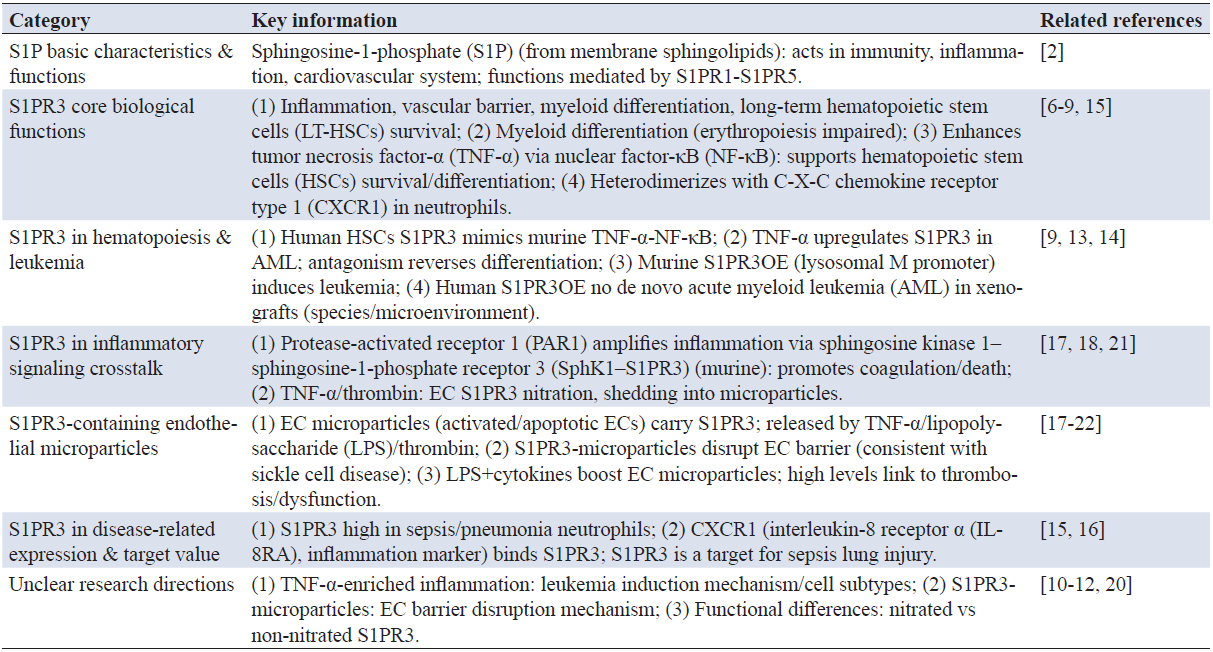
Sepsis is a life-threatening systemic inflammatory response syndrome triggered by infection, characterized by a dysregulated host immune response that often leads to progressive immune dysfunction, organ damage, and even death [1]. Sphingosine-1-phosphate receptors (S1PRs) are an important type of non-Toll-like receptors (non-TLRs) belonging to the G protein-coupled receptor (GPCR) family. They bind to ligands like sphingosine-1-phosphate (S1P), initiating intracellular transmembrane signal transduction, promoting activation of G protein downstream signaling pathways, and enabling diverse biological functions, including regulation of cell proliferation, inflammatory responses, and angiogenesis [2].
Five S1PR subtypes (S1PR1 to S1PR5) have been identified, among which S1PR1, S1PR2, and S1PR3 are ubiquitously expressed. In recent years, S1PR3 has garnered increasing research attention. It is highly expressed in macrophages and can couple with three types of G proteins (Gi, Gq, and G12/13), participating in various pathophysiological processes by regulating oxidative stress responses [3, 4]. Specifically, S1PR3 activates downstream signaling pathways, including PI3K/Akt, phospholipase C (PLC), Rho family GTPases (Rac and Rho), adenylyl cyclase, Jun N-terminal kinase (JNK), and mitogen-activated protein kinase/extracellular signal-regulated kinase (MAPK/ERK), thereby exerting its biological effects [5].
S1P is a bioactive signaling molecule derived from mammalian membrane sphingolipids, functioning in immune and inflammatory responses as well as the cardiovascular system [2]. Most of S1P’s biological functions are mediated by S1PR1 to S1PR5. Among these, S1PR3 participates in numerous biological activities, including inflammation and vascular barrier function [6-8]. S1PR3 promotes myeloid differentiation at the expense of other cell lineages (especially erythropoiesis) and enhances the survival of long-term hematopoietic stem cells (LT-HSCs), closely resembling tumor necrosis factor alpha (TNF-α)-induced emergency hematopoiesis [9]. Thus, S1PR3 acts as an enhancer of TNF-α, utilizing the NF-κB inflammatory signaling pathway to promote HSC survival and myeloid differentiation.
Although TNF-α levels increase with aging and in myeloid malignancies, the mechanisms by which this inflammatory environment induces leukemia and the specific cell subtypes prone to transformation remain unknown [10-12]. Recent studies have revealed that acute TNF-α treatment upregulates S1PR3 membrane levels in primary acute myeloid leukemia (AML) cell lines, while S1PR3 antagonism counteracts TNF-α-induced myeloid differentiation [13]. Previous research demonstrated that S1PR3 overexpression (S1PR3OE) driven by the lysosomal M promoter—rather than the F4/80 promoter—induced leukemic transformation in mice, suggesting that the initiating cell originates from primitive subsets rather than the mature myeloid lineage [13]. However, S1PR3OE in human HSCs did not cause de novo AML in xenografts, indicating that other factors such as the microenvironment and species differences must be considered [14].
S1PR3 expression is elevated in circulating neutrophils of patients with sepsis and pneumonia, forming heterodimers with chemokine (C-X-C motif) receptor 1 (CXCR1), a biomarker of inflammation also known as interleukin-8 (IL-8) receptor alpha (IL-8RA) [15, 16]. This positions S1PR3 as a molecular target for sepsis-associated lung injury.
Furthermore, in vivo studies on S1PR3-mutant mice revealed that protease-activated receptor 1 (PAR1) amplifies inflammation through sphingosine kinase 1 (SphK1)-S1PR3 signaling crosstalk, promoting disseminated intravascular coagulation and death [3]. Endothelial microparticles—complex vesicular structures shed by activated or apoptotic endothelial cells (ECs)—contain enzymes, transcription factors, and mRNA, and are released in response to inflammatory stimuli such as TNF-α, other cytokines, lipopolysaccharide (LPS), and thrombin [17-19]. Studies have shown that EC-damaging substances may induce nitration and shedding of EC S1PR3 into microparticles [20]. Our experiments further demonstrated that S1PR3-containing microparticles enhance EC barrier disruption in vitro, consistent with their role in sickle cell disease, suggesting that microparticles serve as both biomarkers of EC damage and cellular sources of inducers for vascular dysfunction [20].
Combined LPS and cytokine treatment increases EC microparticles production [21]. Microparticles are detectable in plasma of healthy individuals, and higher concentrations under pathological conditions are linked to increased thrombotic risk and endothelial dysfunction (ED) [22]. Related signaling pathways are summarized in Table 1.

3.1 Hepatic fibros
Hepatic fibros is a common consequence of various liver injuries, including viral infection, drug abuse, and alcohol intoxication. After liver injury, levels of SphK1 and S1P increase substantially. In vitro studies have shown that S1PR1/3 mediate S1P-triggered migration of human HSCs and activation of fibrosis [23]. Blockade of S1PR1/3 significantly reduces pathological angiogenesis and hepatic fibrosis in mice with bile duct ligation (BDL) [24]. As hepatic fibrosis progresses, S1P promotes bone marrow mesenchymal stem cell (BMSC) migration via S1PR3. Suramin markedly reduces the number of bone marrow-derived cells during cholestasis, suggesting that the S1P/S1PR3 signaling pathway significantly affects cholestatic hepatic fibrosis by facilitating bone marrow cell homing [25]. Additionally, S1PR1 and S1PR3 are essential for transforming growth factor-β1 (TGF-β1)-induced transdifferentiation of BMSCs into hepatic myofibroblasts [26].
The RNA-binding protein HuR regulates tumor cell migration. HuR mRNA levels are elevated in hepatic fibrotic tissues from humans and mice. In vitro experiments confirmed that the S1PR3/p38 signaling pathway promotes BMSC migration by inducing HuR phosphorylation and cytoplasmic translocation [27]. Furthermore, the S1PR2/3/Gi/PI3K/Rac1 signaling pathway participates in the migration and recruitment of bone marrow mesenchymal monocytes. Administration of the S1PR2 antagonist JTE-013 or the S1PR3 antagonist CAY-10444 significantly reduces recruitment of these monocytes to fibrotic livers, alleviating inflammation and fibrosis in BDL mice [28]. Palmitic acid induces S1P formation in hepatocellular carcinoma cells and promotes its extracellular release. S1P participates in HSC fibrosis via S1PR3. Conditioned medium from hepatocellular carcinoma cells upregulates α-smooth muscle actin (α-SMA) expression in HSCs in an S1P-dependent manner, initiating hepatic fibrosis [29]. Recent studies have shown elevated S1PR3 levels in patients with endometriosis, and S1PR3 participates in TGF-β1-induced fibrosis of endometrial adenocarcinoma cells [29, 30].
Our study provides spatial (zonal) characterization of liver sinusoidal endothelial cells (LSECs; zones 1–3), identification of transcriptomic changes associated with liver cirrhosis, and demonstration of relationships between these transcriptomic changes and phenotypic alterations. Zone 3 LSECs are most susceptible to cirrhosis-associated damage, exhibiting increased capillarization and decreased endocytic capacity. Identification of the most dysfunctional LSEC populations will be valuable for developing effective therapeutic strategies. Furthermore, CD34 is a more useful marker of LSEC capillarization in liver cirrhosis than CD31 [31].
With recent advances in molecular and cell biology technologies, understanding of the pathogenesis of portal vein thrombosis (PVT) associated with liver cirrhosis has deepened. PVT occurrence involves multiple factors, including local and systemic inflammatory responses, disturbances in coagulation-fibrinolysis balance, endothelial dysfunction, and alterations in gut flora [32-34]. The crosstalk among these molecular and cellular mechanisms renders PVT pathogenesis a complex process.
Under cirrhotic conditions, changes in the hepatic microenvironment form an intricate network with systemic immune disorders and coagulation system imbalances. Elucidating the functional network underlying PVT occurrence not only clarifies its pathogenesis but also informs early diagnosis and personalized treatment. This article reviews literature from the past five years, discussing relationships between key factors (inflammatory response, coagulation-fibrinolysis balance, endothelial dysfunction, gut flora) and PVT formation in cirrhotic patients, aiming to provide theoretical support for improving patient management and prognosis [35].
3.2 Regulatory role of S1P and its receptors in sepsis-induced liver injury
S1P and its receptors play key roles in immune and metabo-lic processes, closely associated with various inflammatory responses, oxidative stress, and lipid deposition diseases. As an integral organ for immunity and metabolism, the liver not only defends against bacterial toxins during sepsis but also regulates metabolism of deposited substances post-injury. S1P and S1PR1 to S1PR3 are highly expressed in the liver and act through multiple signaling pathways. For instance, the spinster homolog 2 (Spns2)/S1P signaling pathway exerts inhibitory effects during early inflammatory responses to prevent cytokine storms and promotes later-stage effects to reduce immunosuppression-induced damage [36]. Additionally, S1P likely plays an essential role in bile acid metabolism regulation, particularly relevant in liver injury. Further research on mutual regulation among liver injury, immunity, and metabolism may facilitate discovery of novel therapeutic targets [37]. S1PR-targeted drugs have shown efficacy in regulating immunity, and future studies should explore new metabolic targets to advance treatment strategies for inflammatory and metabolic disturbances associated with sepsis-induced liver injury [38].
3.3 Expression of markers and functional specificity of LSECs versus portal vein endothelial cells
Endothelial cell cultures are valuable tools for investigating liver physiology and pathophysiology in vitro. However, the marked heterogeneity of endothelial cells within and between organs, combined with the tendency for cultured cells to lose tissue-specific markers, complicates such studies. Although all endothelial cells share certain characteristic features, specific markers or marker combinations are needed to define distinct endothelial populations. To date, the study of human sinusoidal endothelial cells (HSECs) in vitro has been hampered by the lack of specific markers that conclusively identify these cells and discriminate them from vascular or lymphatic endothelial cells. This remains partially true, as many classic endothelial markers are widely expressed, and considerable variability exists in phenotypic marker detection between animal and human systems (e.g., CD31 and von Willebrand factor). Currently, no single molecule is known to be expressed exclusively on hepatic sinusoidal endothelium. However, increasing knowledge of endothelial receptors is providing a larger, better-defined set of phenotypic markers. In addition to their utility in phenotyping or sorting specific endothelial cells for culture, tissue-specific receptors offer clues to the functions of the cells under study, exemplified by the numerous scavenger receptors expressed on HSECs.
Very low-passage cells retain fenestrations in vitro for a short time, but these rapidly disappear within one or two passages, as does expression of vascular adhesion protein-1 (VAP-1) [39-41]. Apart from these changes, the cells remain relatively phenotypically stable for 7–8 passages and can be identified by expression of CD31, lymphatic vessel endothelial hyaluronan receptor 1 (LYVE-1), dendritic cell-specific intercellular adhesion molecule-3-grabbing non-integrin-related (L-SIGN), Stabilin-1, and lack of CD34 and prospero homeobox 1 (PROX-1) [39, 42]. These markers confirm endothelial identity while excluding vascular and lymphatic endothelial contamination and, together with markers excluding leukocyte origin, can confirm the sinusoidal nature of the cells [38].
Cardiovascular diseases are leading causes of death worldwide. Endothelial cells, vascular smooth muscle cells, cardiomyocytes, and cardiac fibroblasts all express S1PR1–S1PR3, but expression patterns differ significantly. Endothelial cells primarily express S1PR1 and S1PR3, with S1PR1 being most abundant. Vascular smooth muscle cells highly express S1PR2 and S1PR3. Cardiomyocytes predominantly express S1PR1, followed by S1PR3 at approximately half the level. Cardiac fibroblasts express S1PR3 at much higher levels than S1PR1 and S1PR2 [43].
4.1 Regulation of angiogenesis
In vitro experiments demonstrated that S1P triggers activation of the S1PR3/PI3K/Akt signaling pathway. Under low-concentration S1P stimulation (100 nM), S1PR3 in endothelial progenitor cells primarily mediates Gi protein-dependent PI3K/Akt pathway activation, thereby promoting cell proliferation and inhibiting apoptosis [45].
High-density lipoprotein (HDL)/S1P activates vascular endothelial growth factor receptor 2 (VEGFR2) through S1PR3, promoting proliferation of human umbilical vein endothelial cells and angiogenesis [46]. Li et al. found that S1P/S1PR3 facilitates transdifferentiation of epicardial progenitor cells into smooth muscle-like cells [47]. Recently, Yasuda et al. demonstrated that S1P/S1PR3 promotes corneal angiogenesis by increasing vascular endothelial growth factor A (VEGF-A) production in mice [48].
4.2 Regulation of vascular barrier function
S1PR3 is highly expressed in patients with acute lung injury and severe falciparum malaria complicated by pulmonary edema, primarily in pulmonary endothelial cells and plasma, serving as a potential biomarker for acute lung injury [49]. In LPS-induced acute inflammatory lung injury mouse models, Sammani et al. found that intratracheal administration of high-dose S1P (final plasma concentration ~1 mM) significantly disrupted the alveolar-capillary barrier, an effect prevented by S1PR3 silencing [50]. Different S1P concentrations exert opposing effects on the endothelial barrier: physiological concentrations (10 nM–2 μM) protect the endothelial barrier via the Gi/Rac signaling pathway, whereas higher concentrations activate the S1PR3/RhoA/ROCK pathway, increasing vascular permeability—an effect that may completely counteract the barrier-protective Gi/Rac pathway [51, 52]. Therefore, reducing S1PR3 expression and activation may be an effective strategy for preventing inflammation, acute lung injury, and other diseases linked to endothelial barrier dysfunction [53].
4.3 Regulation of vascular tone
Activating S1PR3 in endothelial cells promotes nitric oxide (NO) production and induces vasorelaxation. In contrast, S1PR3 in vascular smooth muscle cells induces vasoconstriction by increasing intracellular calcium concentration and activating Rho [54]. HDL/S1P promotes calcium mobilization and Gi/PI3K/Akt signaling activation in endothelial cells through S1PR3, facilitating endothelial nitric oxide synthase (eNOS) phosphorylation and ultimately leading to vasodilation [55, 56]. However, other studies have shown that S1P causes vasoconstriction in rat coronary arteries and mouse mesenteric arteries, an effect significantly reduced by pretreatment with the S1PR3-specific antagonist TY52156 [57, 58]. Thus, the effect of S1PR3 activation on vascular tone is complex and requires further detailed investigation.
4.4 Ischemia-reperfusion injury
Accumulating evidence indicates that S1PR3 signaling plays a key role in protecting the heart from ischemia-reperfusion injury (IRI). Administration of HDL/S1P or pretreatment with a specific S1PR3 agonist reduces myocardial infarct size following IRI. Both in vitro and in vivo experiments demonstrate that S1PR3-mediated Akt- and NO-dependent pathways protect cardiomyocytes from apoptosis [52-54]. RhoA has also been implicated in cardioprotection. The G12/13/RhoA/ROCK signaling pathway triggers MRTF-A activation and increases stromal cell protein CCN1 expression, thereby protecting cardiomyocytes from ischemic injury [59-63]. Connexin 43 (Cx43) is a major component of ventricular gap junctions, which transmit action potentials and bioactive substances. Recent studies have shown that S1P regulates Cx43 phosphorylation, expression, and distribution via S1PR2/3, ultimately reducing myocardial cell death and sudden cardiac death caused by IRI [64]. Cx43 uncouplers (e.g., heptanol and Gap26) lower IRI-induced mortality mediated by S1PR2/3 [65]. Interestingly, Wafa et al. found that in the early stage of acute coronary syndrome, S1P/S1PR3 induces coronary artery constriction and impairs myocardial contractile function, although S1PR3-induced vasoconstriction may be counteracted by S1PR2-mediated vasorelaxation [53-66]. Thus, S1PR3 activation may represent an important strategy for preventing myocardial IRI.
Studies have demonstrated that S1PR3 participates in inflammation, proliferation, migration, tumor invasion, IRI, tissue fibrosis, and vascular activity [67]. In a mouse model of myocardial IRI, HDL and its component S1P protect the heart via an S1PR3-mediated signaling pathway [68]. S1PR3(–/–) mice are protected from renal IRI through mechanisms involving bone marrow-derived dendritic cells (BMDCs) and their immunomodulatory functions [69]. Adoptive transfer of S1PR3(–/–) BMDCs prevents renal IRI through splenic interactions and expansion of splenic CD4+Foxp3+ regulatory T cells. In contrast to its protective role in cardiac IRI, S1PR3 exerts opposite effects in renal IRI, likely due to differences in disease stage and cell/tissue contexts.
S1PR1 and S1PR2 have been implicated in microglial activation during cerebral IRI [70-72]. A recent study showed that S1PR3 contributes to microglial activation and M1 macrophage polarization in a mouse brain IRI model [73]. Our study demonstrated that S1PR3 inhibition reduces IRI, confirmed by reduced infarct volume. H&E, Nissl, and Fluoro-Jade C staining confirmed that S1PR3 mediates brain damage during cerebral IRI [74].
Neuronal nitric oxide synthase (nNOS) mediates early neurological damage, with nNOS overexpression playing a key role in early ischemia and excitotoxic injury [75]. Inducible NOS (iNOS) subsequently increases, and both adversely affect cerebral ischemia. NO production, catalyzed by NOS from L-arginine and oxygen, can react with superoxide to form peroxynitrite, a potent oxidant [76]. During ischemia, NO produced by nNOS and iNOS may be neurotoxic, partly due to peroxynitrite free radical formation causing direct damage to mitochondrial enzymes and DNA [77]. Additionally, increased NO production from nNOS or iNOS promotes ischemic damage through free radical injury, tissue inflammation, and microcirculatory failure [78]. nNOS knockout mice exhibit smaller infarct sizes and fewer neurological deficits after middle cerebral artery occlusion [79]. iNOS knockout mice also show fewer neurological deficits and reduced infarct volumes [80]. In our experiment, nNOS and iNOS expressions were elevated following cerebral ischemia compared with sham-operated controls, consistent with previous studies. S1PR3 inhibition reduced nNOS expression and NO content. Heo and Im found that S1PR3 inhibition reduces LPS-induced inflammatory gene expression, including iNOS and cyclooxygenase-2 [74]. However, our study found that S1PR3 inhibition did not reduce iNOS expression, suggesting that S1PR3 reduces NO primarily by decreasing nNOS rather than iNOS.
4.5 Atherosclerosis
Atherosclerosis is a chronic inflammatory disease, with ED being a key driver of its development. HDL is considered atheroprotective. Kimura et al. found that HDL activates PI3K/Akt, p38, and Rho/ROCK pathways via S1P/S1PR3, promoting endothelial cell migration and exerting anti-atherosclerotic effects [81]. Additionally, S1P prevents macrophage apoptosis by activating the S1PR3/STAT3/surviving signaling pathway, reducing the risk of plaque rupture in advanced atherosclerotic lesions [82].
Macrophages with S1PR3 knockout are resistant to 22(R)-hydroxycholesterol-stimulated cholesterol efflux. Conversely, the S1P/S1PR3/Gq/PLC/protein kinase C (PKC) signaling pathway may promote cholesterol efflux by phosphorylating and stabilizing the ATP-binding cassette transporter A1 (ABCA1) protein [83]. A recent study emphasized the core role of ED as a major precursor of inflammation, not only accelerating atherosclerosis progression but also causing plaque instability. Hallmarks of ED include reduced NO production/availability and endothelial cells exhibiting an inflammatory phenotype. The endothelial glycocalyx, a gel-like structure composed of glycoproteins and extracellular matrix components, participates in transendothelial transport. Endothelial glycocalyx damage impairs eNOS expression, leading to reduced NO availability and production [84].
Various inflammatory signals activate endothelial cells, triggering upregulation of cell adhesion molecules, including pro-inflammatory E-selectin, intercellular adhesion molecule-1 (ICAM-1), and vascular cell adhesion molecule-1 (VCAM-1). Leukocyte diapedesis occurs, accompanied by increased endothelial permeability [85]. Activated endothelial cells secrete cytokines and function similarly to macrophages. Dysfunctional, inflamed endothelium facilitates lipoprotein penetration and trapping beneath the endothelial cell layer, where these lipoproteins are subsequently oxidized [66]. Deposited lipoproteins are taken up by macrophages and smooth muscle cells, leading to foam cell formation and fatty streak accumulation. Abnormalities in cell clearance—specifically, failure to properly remove apoptotic endothelial cells—result in vascular sclerosis [86]. Some endothelial cells may undergo endothelial-mesenchymal transition, promoting extracellular matrix deposition and further exacerbating vascular stiffness [87]. Arterial constriction caused by lipid accumulation, combined with impaired vasodilation, expands regions of turbulent or oscillatory blood flow, further intensifying endothelial cell activation and denudation. These regions carry a high risk of rupture for lipid-rich plaques with thin fibrous caps, potentially leading to thrombotic occlusion. Expression of procoagulant factors—tissue factor, plasminogen activator inhibitor-1 (PAI-1), and von Willebrand factor (vWF)—further heightens this risk [84-88]. Thus, ED serves as a precursor to both atherosclerosis and thrombosis.
Patients with coronary artery disease often experience sleep disturbances, reduced melatonin levels, and circadian rhythm misalignment [89]. Sleep disturbances are linked to higher levels of inflammatory markers, ED, and reduced cardiac vagal modulation—all contributing to coronary artery disease development and progression [90]. Relatively few studies have established a clear connection between lifestyle behaviors (e.g., physical activity level, a modifiable cardiovascular risk factor) and endothelial function in patients with cardiovascular disease [91]. In the present study, multiple regression analysis revealed a significant association between low physical activity levels and ED (Table 1). Analysis of the control group found that physically active patients had better endothelial function than sedentary ones. Consistent with this, a study involving 2,363 patients with prediabetes and type 2 diabetes showed that sedentary behavior is linked to higher levels of low-grade inflammatory biomarkers (C-reactive protein, soluble ICAM-1, IL-6, and TNF-α) as well as biomarkers of ED, which are key components of the atherosclerotic process illustrated in Figure 1 [92].
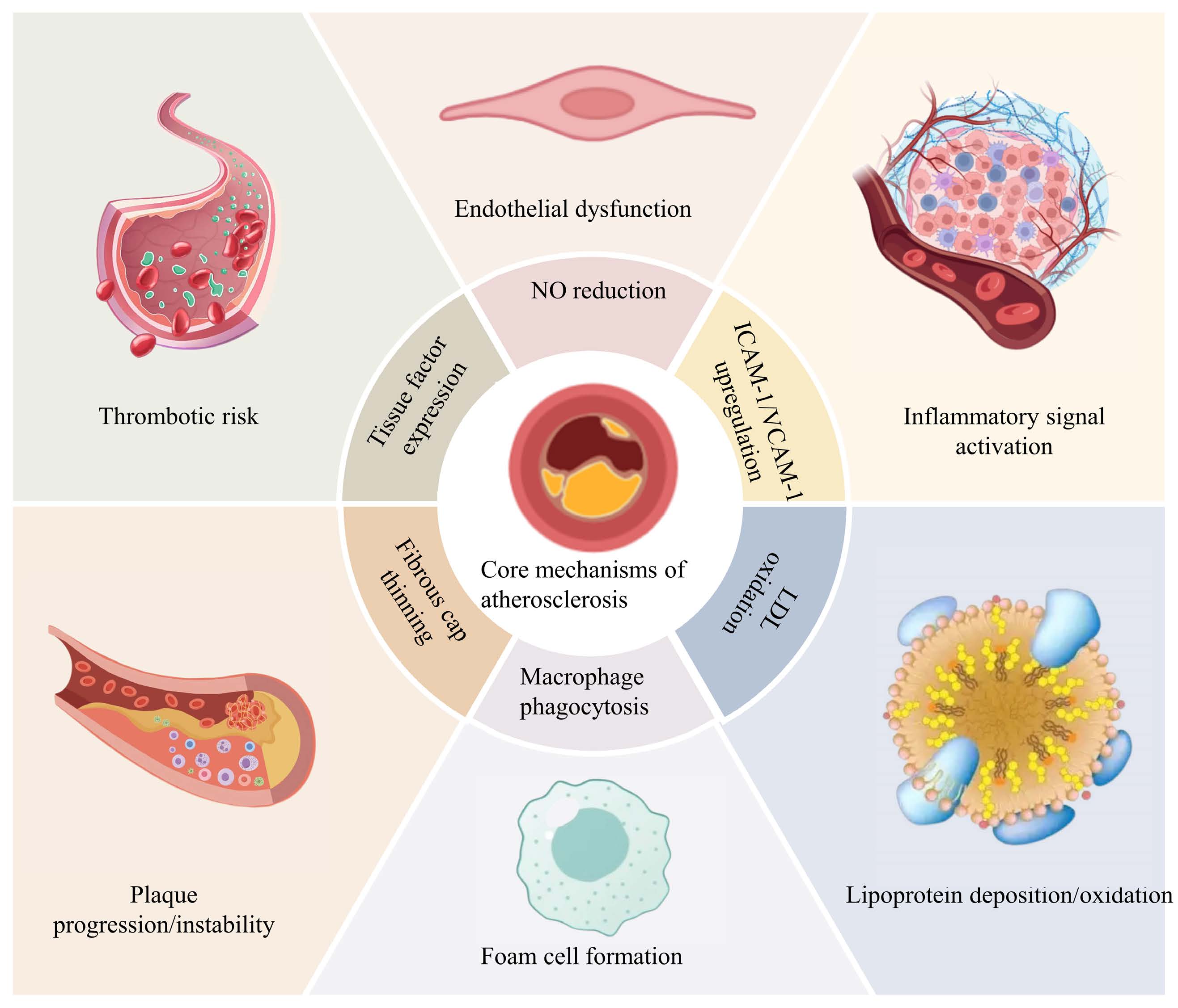
Figure 1. Core mechanisms of atherosclerosis. Binding of sphingosine-1-phosphate (S1P) to S1P receptor 3 (S1PR3) exerts pleiotropic effects in vascular biology, regulating angiogenesis, vascular tone, atherosclerosis, and ischemia-reperfusion injury protection via distinct signaling pathways. This figure depicts the core mechanisms of atherosclerosis, including endothelial dysfunction (manifested as reduced nitric oxide (NO) bioavailability and upregulated intercellular adhesion molecule 1 (ICAM-1)/vascular cell adhesion molecule 1 (VCAM-1)), inflammatory signaling activation, lipoprotein deposition and oxidation (involving low-density lipoprotein (LDL) oxidation), macrophage phagocytosis and subsequent foam cell formation, plaque progression and instability (characterized by fibrous cap thinning and tissue factor expression), as well as heightened thrombotic risk. Created with Microsoft PowerPoint.
Growing evidence suggests that S1PR3 regulates the biological functions of certain immune cells, including dendritic cells (DCs), macrophages, and natural killer cells. In vitro studies have shown that S1PR3 is essential for S1P-induced maturation, migration, and endocytosis of DCs [69]. Additionally, S1PR3 in DCs plays a key role in recruiting NKT cells and neutrophils to damaged kidneys following IRI. Notably, DCs lacking S1PR3 induce a Th2-like response in NKT cells—specifically prompting them to produce higher levels of IL-4—which protects the kidneys against IRI [69, 93].
S1PR3 is a pro-inflammatory receptor. Hyperglycemia inhibits M2 macrophage polarization and promotes M1 polarization via the S1P/S1PR3 signaling pathway, worsening hepatic IRI. However, pretreatment of diabetic mice with CAY10444 (an S1PR3 antagonist) significantly reduces hepatic IRI [94].
S1PR2 and S1PR3 promote migration of bone marrow-derived monocytes/macrophages by activating the Gi/o/PI3K/Rac1 signaling pathway. Administration of JTE-013 (S1PR2 antagonist) or CAY-10444 (S1PR3 antagonist) to BDL-treated mice significantly reduces recruitment of these cells to fibrotic livers [4, 26]. Yang et al. found that S1PR2/3 activates the Gi/PI3K/JNK signaling pathway, mediating S1P-induced M1 polarization of bone marrow-derived monocytes/macrophages [95]. However, Bryan et al. showed that FTY720 reduces macrophage phagocytic capacity and reactive oxygen species production via S1PR3, thereby reactivating Cryptococcus infection in granulomas [96]. Therefore, S1PR3 exerts dual roles in macrophages: promoting M1 polarization to trigger inflammation while also enhancing phagocytic and killing activities to inhibit bacterial diseases. Figure 2 provides a schematic overview of these roles.
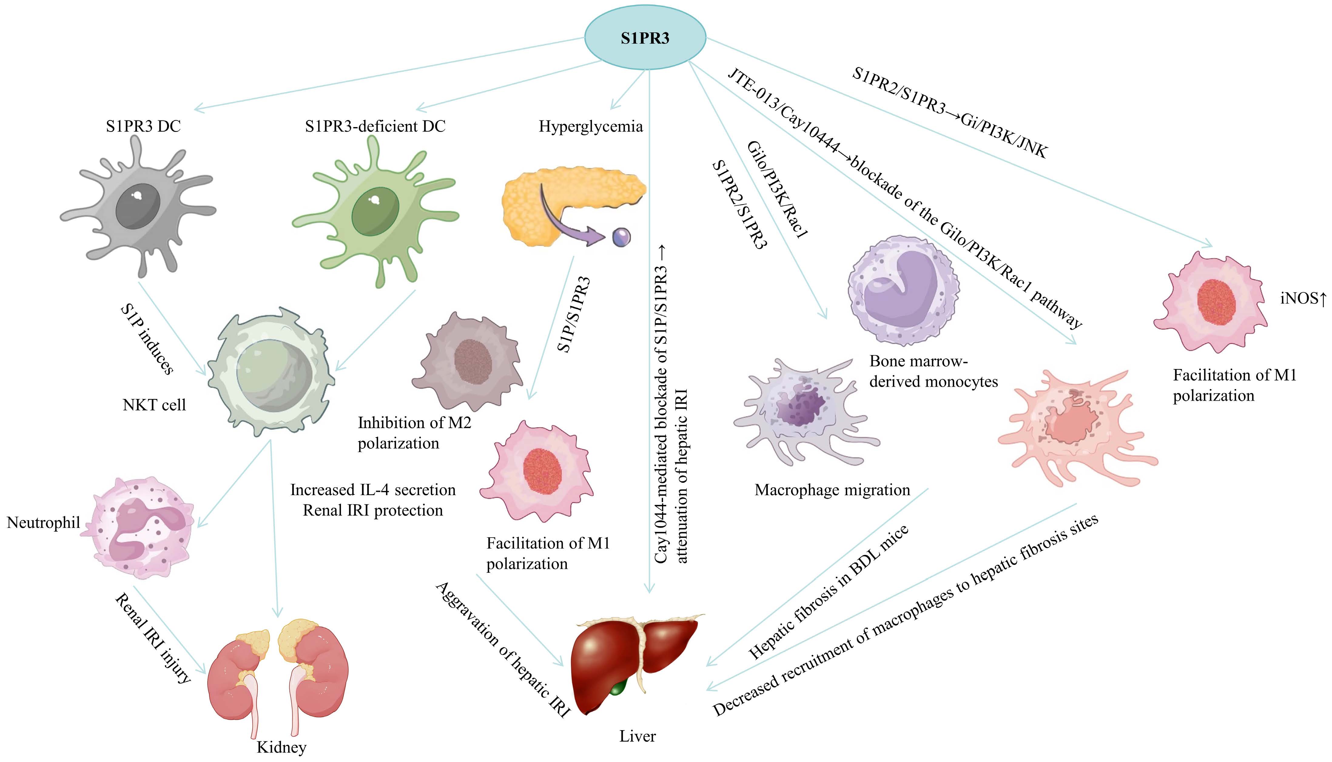
Figure 2. Multifaceted roles of S1PR3 in immune cell regulation and tissue injury responses. S1PR3 orchestrates immune cell functions, including those of dendritic cells (DCs) and macrophages, to mediate renal ischemia-reperfusion injury (IRI), hepatic injury/fibrosis, and antiinfection responses. This figure outlines the multifaceted roles of S1PR3. In renal IRI, S1PR3-expressing DCs (S1PR3 DCs) trigger natural killer T (NKT) cell activation and interleukin-4 (IL-4) secretion, whereas S1PR3-deficient DCs inhibit M2 macrophage polarization, together shaping renal injury outcomes; in hepatic IRI and fibrosis, S1PR3 signals through Gi/o/PI3K/Rac1 or Gi/PI3K/JNK pathways to drive M1 macrophage polarization (marked by elevated inducible nitric oxide synthase (iNOS) expression) and macrophage migration, while S1PR3 blockade with CAY10444 alleviates hepatic IRI and reduces macrophage recruitment to fibrotic lesions; urthermore, hyperglycemia and S1P/S1PR3 signaling amplify M1 polarization to worsen hepatic IRI, and S1PR3 promotes the migration of bone marrow-derived monocytes. Created with Microsoft PowerPoint.
Astrocytes are one of the most common cell types in the brain and are critical for maintaining the brain’s microenvironment [97]. In vitro studies have demonstrated abundant S1PR3 expression in astrocytes, which increases further during inflammation. S1PR3-G12/13 activates RhoA, leading to increased production of inflammation-related genes [98]. CAY10444, a specific S1PR3 antagonist, has shown protective effects in models of cerebral infarction, spinal cord injury, and intracerebral hemorrhage [99-101].
In vitro experiments show that S1P/S1PR3/Ras/pERK signaling promotes pericyte proliferation, contributing to scar formation after spinal cord injury [99]. Dong et al. found that FTY720P protects astrocytes from oxygen-glucose deprivation-induced injury and inflammation by inhibiting S1PR3-dependent TLR2/4-PI3K-NFκB signaling [101]. Additionally, S1PR3 inhibition exerts neuroprotective effects via the S1P/CCL2/p-p38 MAPK pathway [100]. S1PR3 antagonists may represent a potential therapeutic approach for nervous system injuries.
ED is a hallmark of ARDS [99]. The endothelium is a monolayer of cells that controls the permeability of fluids, proteins and blood cells [86]. In ARDS, endothelial barrier permeability increases as monolayer integrity is disrupted, allowing fluid to enter interstitial tissue, further decreasing gas exchange and inducing hypoxia [102, 103]. Endothelial cell-cell adhesions, including adherens junctions (AJs), tight junctions, and gap junctions, regulate barrier permeability [104]. Among these, AJs have more significant microvascular effects, and expression of vascular endothelial cadherin (VE-cadherin) at endothelial AJs is essential for endothelial barrier function [48]. AJ structure involves interactions among VE-cadherin, β-catenin, and the actin cytoskeleton [105]. In this study, we found that in LPS-induced ARDS mouse models and LPS-stimulated cell models, S1PR3 inhibition significantly restored expression of VE-cadherin and β-catenin, as shown by Western blotting and immunofluorescence [106]. Additionally, rhodamine phalloidin staining of the cytoskeleton revealed that S1PR3 inhibition significantly reduced LPS-induced cytoskeletal disruption. S1PR3 mediates VE-cadherin mRNA levels in two types of endothelial cells. In a murine ventilator-induced lung injury model, S1PR3 induced endothelial barrier loss through ADAM10-mediated VE-cadherin cleavage [107].
To date, more than 600 clinical trials have targeted Rho signaling [108-110]. In contrast, no clinical trial targeting S1PR3 has ever been conducted. Targeting S1PR3 in clinical trials may be beneficial, as S1PR3 is not only an upstream regulator of Rho but also a mediator of other pathways, such as VE-cadherin cleavage and NFκB-mediated inflammation. Moreover, most existing S1P-modulated drugs targeting S1PR1 agonistically induce downregulation of S1PR1, leading to suboptimal clinical effects and long-term pulmonary function decompensation due to immunosuppression [111]. In contrast, inhibiting S1PR3—a negative regulator of vascular barrier function upregulated during mechanical stress—does not affect S1PR1 expression, ensuring specific therapeutic effects during ventilator-induced lung injury with few side effects under normal lung conditions. However, S1PR3 is also expressed in the cardiovascular system, kidney, and spleen [112, 113]. S1PR3 inhibition could also lead to hypertension, bradycardia, macular edema, reduced pulmonary function, hepatic adverse effects, and neoplasms [114]. Inhalation delivery of S1PR3 inhibitors via aerosol or nanoparticles, which facilitates direct delivery of drugs deep into the airways, may mitigate these effects. Notably, TY-52156, although a selective S1PR3 inhibitor, may carry significant off-target effects, including inhibiting other S1P receptors with relatively lower affinity [115].
S1P, a metabolic mediator of sphingolipid catabolism predominantly formed through SphK1 catalysis, mediates inflammation in sepsis by binding to S1PR3 expressed in macrophages. We demonstrated that the SphK1/S1PR3 axis is upregulated in LPS-induced macrophages and septic mouse lungs, cascading activation of pro-glycolytic signaling molecules such as HIF-1α, HK2, and PFKFB3. Targeted inhibition of SphK1 by PF-543 effectively abrogated the upregulated SphK1/S1PR3 axis in vitro and in vivo. Additionally, PF-543 significantly suppressed sepsis-related inflammation and multi-organ injury in vivo. Furthermore, PF-543 not only blunted key glycolytic enzymes (HIF-1α, HK2, and PFKFB3) in LPS-treated macrophages but also inhibited HK2 and PFKFB3 in septic mice. Silencing or inhibiting SphK1 tempered pro-inflammatory M1 macrophages while boosting anti-inflammatory M2 macrophages. Intriguingly, S1PR3 knockdown efficiently dampened glycolysis-associated markers, reversed LPS-modulated M1/M2 polarization, and attenuated NF-κB p65 activation. In conclusion, this study provides the first evidence that PF-543 orchestrates proportional imbalance of macrophage polarization and the Warburg effect in a SphK1/S1PR3-dependent manner during sepsis, mitigating both hyperinflammation and multi-organ failure, adding a novel puzzle piece to pharmacologically exploitable therapy for sepsis [116].
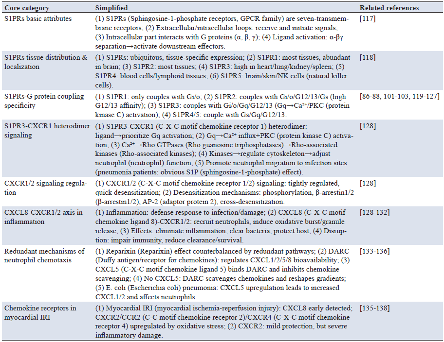
Sphingosine-1-phosphate receptors are members of the GPCR family, also known as seven-transmembrane receptors. The peptide chains that traverse the membrane seven times form extracellular and intracellular loops, receiving extracellular signals and initiating intracellular signaling. The intracellular domain interacts with heterotrimeric G proteins, composed of α, β, and γ subunits. Upon ligand activation, the receptor binds G proteins, causing dissociation of the α subunit from the βγ subunits, which then activate downstream effector molecules and regulate biological effects [117].
S1PRs are ubiquitously expressed but exhibit tissue-specific patterns. S1PR1 and S1PR2 are expressed in most tissues: S1PR1 localizes to the plasma membrane, vesicles, cytoplasm, nucleus, and perinuclear regions, while S1PR2 is found in the plasma membrane and cytoplasm. S1PR3 is highly expressed in heart, lung, kidney, and spleen, and is located in the plasma membrane; S1PR4 is mainly expressed in blood cells and lymphoid tissues; S1PR5 is expressed in brain, skin, and natural killer cells [118].
S1PR1 is the most widely expressed S1P receptor in the brain and couples only with Gi/o [83-85]. S1PR2 can couple with Gi/o, G12/13, and Gs, exhibiting highest binding affinity for G12/13 [103, 104, 117-120]. S1PR3 is known to couple with Gi/o, Gq, and G12/13; however, due to its higher affinity for Gq, it can induce intracellular calcium accumulation and PKC activation [88-90]. S1PR4 and S1PR5 can couple with Gs, Gq, and G12/13 [121-127].
S1PR3 has relatively high affinity for Gq proteins. When it forms a heterodimer with CXCR1, ligand binding still prioritizes Gq pathway activation, driving extracellular calcium influx and simultaneously switching on PKC. Once the calcium signal is activated, it further triggers activation of Rho family small GTPases, which then activate downstream Rho-associated protein kinases, regulating cytoskeletal assembly and disassembly by phosphorylating substrates such as myosin light chains. This process adjusts neutrophil morphology and motility, facilitating their migration across the vascular wall to infection sites. Interestingly, this regulatory effect is only significantly evident in neutrophils from pneumonia patients and is induced by S1P. CXCR1/2 G-protein signaling is tightly regulated and quickly desensitized to prevent constitutive signaling through mechanisms including phosphorylation, β-arrestin1/2 recruitment, AP-2 adaptor protein association, and receptor cross-desensitization.Inflammation is a defense mechanism triggered by infection and tissue damage [128]. The CXCL8-CXCR1/2 axis recruits neutrophils to infection sites and induces neutrophil oxidative burst and granule release to eliminate inflammatory stimuli and increase bacterial clearance, thereby protecting the host from further infection and tissue damage [129-131]. Disruption of this axis severely affects host immune mechanisms against infection and may even be fatal. Impaired neutrophil recruitment often leads to decreased bacterial clearance and reduced survival rates in experimental infectious disease models [128].
Given the multiplicity of CXCR1 and CXCR2 ligands and the more selective inhibition of the CXCR1-related pathway via reparixin, the impact of reparixin could be counterbalanced by redundant mechanisms of neutrophil chemotaxis, resulting in perpetuation of inflammation independently of the initial signal. For example, the Duffy antigen receptor for chemokines (DARC), considered a “silent” chemokine receptor, has recently been shown to regulate bioavailability of several chemokines, including CXCL1, CXCL2, CXCL5, and CXCL8, resulting in DARC-enhanced neutrophil trafficking into tissues [132, 133]. The CXCR2 ligand CXCL5 regulates binding site availability for other ELR+ CXC chemokines released during inflammation through its interaction with erythrocyte DARC. CXCL5 inhibits chemokine scavenging, at least in part, through its homeostatic, high-affinity binding with erythrocyte DARC. Thus, in the absence of CXCL5, DARC scavenges proinflammatory chemokines, contributing to reshaping chemokine gradients for neutrophil influx into, for example, the lung [134]. During severe inflammatory responses, such as in a model of Escherichia coli-induced pneumonia, further expression of CXCL5 by alveolar type 2 cells inhibits DARC chemokine scavenging when production of CXCL1 and CXCL2 dramatically increases, resulting in marked increases in circulating plasma concentrations of these chemokines and adverse consequences for efficient neutrophil accumulation [134, 135]. This example illustrates the complexity and alternative pathways of neutrophil recruitment and regulation independent of overwhelming CXCR1 pathway blockade [134].
Cardiac dysfunction has been associated with elevated circulating chemokine levels in both animals and humans. CXCL8 was among the first chemokines detected in the myocardium in response to IRI, and it has been hypothesized to participate in neutrophil-induced myocardial injury. However, several other chemokines, including CCL2 and CXCL10, have been detected early in animal models of myocardial injury. Cardiac myocytes also express other receptors such as CXCR2, CCR2, and CXCR4 (the receptor for SDF-1/CXCL12). These receptors are constitutively expressed and upregulated following oxidative stress both in vivo and in vitro [136]. In an animal model of IRI, Tarzami et al. found a direct myocardial protective action of CXCR2 during IRI, but the magnitude of this protective effect was smaller than the damaging effect of CXCR2-mediated inflammatory cell recruitment [137]. The authors attributed this to different CXCR2-induced signaling between two different cell types—cardiac myocytes and neutrophils [136]. Additionally, new data highlight the relevance of the CXCL12/CXCR4 axis in myocardial IRI. CXCR4 overexpression in vivo worsened cardiac function in an animal model of IRI, enhancing inflammatory cell recruitment, TNF-α production, and cell death, possibly due to increased CXCL12 production [137, 138]. Furthermore, under stress conditions, paracrine and/or autocrine activation of CXCR4 by its ligand may trigger intracellular signaling pathways that exacerbate contractile dysfunction. CXCR4 activation also modulates β2-adrenergic receptor structure and downstream signaling in ventricular cardiac myocytes and has been shown to be a key mediator of calcium handling in adult rat cardiac myocytes [139]. Thus, independently of reparixin action, myocardial IRI may be triggered and maintained by different chemokine/chemokine-receptor inter-relationships (e.g., the CXCL12/CXCR4 axis), which could explain the lack of significant differences in primary endpoints for myocardial IRI in our study [134]. The biological characteristics and functions of S1P and S1PR3 related to these processes are summarized in Table 2.
Note: S1P, Sphingosine-1-phosphate; S1PR3, Sphingosine-1-phosphate receptor 3; LT-HSCs, Long-term hematopoietic stem cells; TNF-α, Tumour necrosis factor-α; NF-κB, Nuclear factor kappa-B; CXCR1, C-X-C chemokine receptor type 1; AML, Acute myeloid leukemia; S1PR3OE, S1PR3 overexpression; PAR1, Protease-activated receptor 1; SphK1, Sphingosine kinase 1; EC, Endothelial cell; LPS, Lipopolysaccharide; IL-8RA, Interleukin-8 receptor alpha.
S1PR3 plays a critical role in regulating inflammatory responses and endothelial barrier function in ARDS. It is highly expressed in lung tissue and endothelial cells and has been widely described as a mediator of various biological responses, but its exact role varies across different cells, tissues, and diseases under distinct conditions [140]. Wang et al. reported that S1PR3 inhibition suppressed NLRP3 inflammasome activation in LPS-stimulated macrophages and in the lungs of septic mice [141]. Bajwa et al. reported that S1PR3-deficient dendritic cells exerted protective effects against IRI-induced acute kidney injury [7]. However, the relationship between S1PR3 and ARDS remains unclear. Our results demonstrated that S1PR3 expression was increased in the lungs of LPS-induced ARDS mice, and S1PR3 inhibition alleviated inflammation and endothelial injury both in vivo and in vitro, thereby reducing LPS-induced ARDS [142].
Mechanistically, S1PR3 modulates key inflammatory signaling cascades, including the NF-κB pathway, to orchestrate the production of pro-inflammatory cytokines such as TNF-α, IL-1β, and IL-6 [142, 143]. The NF-κB pathway is considered the central regulator of inflammation and endothelial function in various diseases, and our previous study revealed the importance of the NF-κB pathway in ARDS [143, 144].
NF-κB has five subunits, with p65 being the key transcriptionally active component. IκBα is a crucial controller of NF-κB p65 activation and is phosphorylated by IKKβ to release active NF-κB p65 into the nucleus, thereby promoting transcription of proinflammatory cytokines and chemokines [145]. In endothelial cells, activated NF-κB contributes to VCAM-1 expression, mediating endothelial inflammation and damage [146]. Previous research has indicated an association between S1PR3 and the NF-κB pathway. Yan et al. reported that the NF-κB pathway participates in S1PR3-mediated human renal cell carcinoma progression [147]. Moreover, previous studies have revealed the NF-κB-dependent proinflammatory role of the S1P/S1PR3 axis in lung adenocarcinoma cells, with S1PR3 activation facilitating tumor cell proliferation [136, 148]. Mitochondria, the main powerhouses of cells, are crucial for regulating inflammatory responses and barrier function in endothelial cells [135]. Mitochondrial oxidative phosphorylation is the primary pathway for ATP production, dependent on mitochondrial membrane potential and respiratory chain complex activity [149]. Consistent with previous studies, we found that LPS suppressed mitochondrial oxidative phosphorylation [150]. S1PR3 inhibition decreased reactive oxygen species levels in lung tissues and mitochondrial ROS levels in endothelial cells while increasing the aggregate-to-monomer ratio. Additionally, ATP production and mitochondrial respiratory chain complex activity were restored after S1PR3 inhibition. The complex V inhibitor oligomycin abolished the protective effect of S1PR3 inhibition on ARDS. Therefore, mitochondrial oxidative phosphorylation may mediate the S1PR3 protective effect in ARDS [142].
Preclinical studies have shown that S1PR1 and S1PR3 may be primary contributors to S1P receptor modulator (S1PRM)-induced cardiovascular adverse events [151]. Due to initial S1PR1 agonism, S1PRM administration triggers a decrease in heart rate and blood pressure, activating G protein-coupled inwardly rectifying potassium channels, followed by myocyte hyperpolarization and transient reduction in excitability. Continuous administration leads to S1PR1 downregulation, subsequent shifts in the S1P receptor profile, and increased blood pressure [152].
Furthermore, S1PR3 agonism may play a role in heart rate effects: fingolimod induces bradycardia in wild-type mice, but not in S1PR3 knockout mice [153, 154]. However, the potential involvement of S1PR3 in humans remains unclear. Although extensive efforts have been made to develop new modulators with lower affinity for S1PR3 (e.g., siponimod and ozanimod), minimizing S1PR3 binding does not prevent the cardiac effects observed upon first-dose administration [155-159].
A recent study employed gene clustering functional analysis to identify signaling pathways affected by CAY10444 treatment and S1PR3 knockdown, aiming to explore molecular pathways through which targeting S1PR3 influences WEE1 expression [160]. Although CAY10444 treatment affected the PI3K/Akt, PI3K/Akt/mTOR, and MAPK signaling pathways, the PI3K/Akt pathway was the only common pathway affected by both treatments. Studies have reported that S1PR3 can bind to various Gα proteins, including Gi, Gq, and G12/13, thereby regulating signaling pathways such as adenylyl cyclase/cAMP, Ras/ERK/AC, PI3K/Akt, PLC/Ca²⁺, and Rho/ROCK [161].
Thus, targeting S1PR3 in oral squamous cell carcinoma (OSCC) may inhibit PI3K/Akt signaling pathway activation. The PI3K/Akt pathway exerts crucial biological functions in tumors [162]. Results confirmed that S1PR3 inhibition decreased Akt phosphorylation levels. Conversely, the Akt agonist SC79 restored S1PR3 inhibition-induced suppression of OSCC cell growth both in vitro and in vivo. Additionally, SC79 partially reversed the downregulation of WEE1 and phosphorylated CDC2 induced by S1PR3 targeting. These findings indicate that S1PR3 knockdown inhibits OSCC cell growth by regulating the Akt/WEE1 signaling pathway.
S1PR3 is a GPCR and seven-transmembrane receptor that binds to extracellular S1P, activating downstream signaling pathways. S1PR3 plays important roles in controlling normal physiological functions and disease processes across multiple systems, including the cardiovascular, immune, nervous, and hepatic systems. S1PR3 exhibits cell- and tissue-specific effects, contributing to both maintenance of normal tissue homeostasis and pathogenesis of diseases such as inflammation, IRI, and atherosclerosis.
This article systematically summarizes the core biological characteristics of S1PR3, its mechanisms of action in various systems, and its prospects for clinical application. It also clarifies the dual roles and regulatory rules of S1PR3 in inflammation, IRI, and atherosclerosis. Notably, conflicting conclusions exist in current research, with the most typical being the controversy over S1PR3’s role in myocardial IRI. Some studies have confirmed that activating S1PR3 inhibits cardiomyocyte apoptosis and reduces infarct size via the Gi-Akt-NO pathway; however, others have found that it exacerbates cell damage via the G12/13-RhoA pathway. The core of this contradiction lies in the failure to fully consider the context-dependent activation characteristics of the S1PR3 signaling pathway.
Based on the mechanistic analysis in this article, this controversy can be explained from three aspects. First, differences in injury stage: in the early stage of ischemia (0~2 h after reperfusion), cardiomyocytes secrete low-concentration S1P (approximately 500 nM) in response to stress. At this time, S1PR3 preferentially couples with highly expressed Gi proteins to initiate the PI3K/Akt protective pathway. In the late stage of reperfusion (>6 h), inflammatory factors induce S1P concentrations to rise above 2 μM and simultaneously upregulate G12/13 protein expression, shifting the pathway toward a pro-injury direction. Differences in intervention timing among different studies directly lead to divergent results. Second, cellular heterogeneity: basal Gi protein expression levels in cardiomyocytes are significantly higher than those in vascular smooth muscle cells, and they retain partial Gi coupling activity even in pathological high-concentration S1P environments. However, some studies have failed to distinguish signal differences between cardiomyocytes and interstitial cells, potentially confounding overall injury effects with cell-specific roles. Third, differences in regulatory methods: non-specific agonists (such as S1P itself) are likely to trigger pathway switching due to concentration fluctuations, whereas specific agonists targeting the S1PR3-Gi coupling domain can stably exert protective effects. This also explains why different intervention strategies yield opposite conclusions.
Currently, several deficiencies remain in S1PR3-related research. Besides the PI3K/Akt, PLC/Ca²⁺, and RhoA/ROCK pathways, crosstalk mechanisms between S1PR3 and other signaling pathways have not been fully elucidated. The specific molecular switches (e.g., specific phosphorylation sites and regulatory proteins) governing S1PR3 signaling pathway conversion under different disease states still need to be identified. Targeted delivery systems and side effect control strategies for S1PR3-specific modulators require optimization. In the future, combining technologies such as single-cell sequencing and proteomics to analyze context-dependent activation mechanisms of the S1PR3 signaling pathway and developing cell-specific S1PR3-modulating drugs will provide new directions for precision treatment of related diseases.
Author contributions
Yichen He, Xiangyi Zhang, and Qin Zhang contributed to the study design and manuscript drafting; Weiqi Lin, Yan Zhang, Haiyi Qian, and Wen Ke were responsible for data acquisition and statistical analysis; Cuifeng Zhang and Qun Chen provided critical insights during data interpretation, participated in constructive discussions, and revised the manuscript for intellectual content.
Funding
This review article was conducted as part of a thesis project funded by Natural Science Foundation of the Higher Education Institutions of Anhui Province (2025AHGXZK30987), The Foundation of Wannan Medical University (WK2023ZQNZ08), National university innovation and entrepreneurship training program (202410368019, 202410368018, 2025103668067), Anhui Province university innovation and entrepreneurship training program (S202410368004, S202410368009, S202310368095, S202210368103), Lunan Houpu Pharmaceutical Co., Ltd. Horizontal Research Project (HXKT2022012).
Data availability
All source data for this work (or generated in this study) are available upon reasonable request.
Ethical approval and consent to participate
Not applicable.
Consent for publication
Not applicable.
Competing interests
The authors declare that they have no competing interests.
Acknowledgements
Not applicable.
[2] Kunkel GT, Maceyka M, Milstein S, Spiegel S. Targeting the sphingosine-1-phosphate axis in cancer, inflammation and beyond. Nat Rev Drug Discov. 2013 Sep;12(9):688-702. https://doi.org/10.1038/nrd4099
[3] Niessen F, Schaffner F, Furlan-Freguia C, Pawlinski R, Bhattacharjee G, Chun J, et al. Dendritic cell PAR1-S1P3 signalling couples coagulation and inflammation. Nature. 2008 Apr 3;452(7187):654-658. https://doi.org/10.1038/nature06663
[4] Keul P, Lucke S, von Wnuck Lipinski K, Bode C, Gräler M, Heusch G, et al. Sphingosine-1-phosphate receptor 3 promotes recruitment of monocyte/macrophages in inflammation and atherosclerosis. Circ Res. 2011 Feb 4;108(3):314-323. https://doi.org/10.1161/CIRCRESAHA.110.235028
[5] Pyne S, Pyne NJ. Sphingosine 1-phosphate signalling in mammalian cells. Biochem J. 2000 Jul 15;349(Pt 2):385-402. https://doi.org/10.1042/0264-6021:3490385
[6] Muppidi JR, Lu E, Cyster JG. The G protein-coupled receptor P2RY8 and follicular dendritic cells promote germinal center confinement of B cells, whereas S1PR3 can contribute to their dissemination. J Exp Med. 2015 Dec 14;212(13):2213-2222. https://doi.org/10.1084/jem.20151250
[7] Bajwa A, Huang L, Kurmaeva E, Gigliotti JC, Ye H, Miller J, et al. Sphingosine 1-phosphate receptor 3-deficient dendritic cells modulate splenic responses to ischemia-reperfusion injury. J Am Soc Nephrol. 2016 Apr;27(4):1076-1090. https://doi.org/10.1681/ASN.2015010095
[8] Nussbaum C, Bannenberg S, Keul P, Gräler MH, Gonçalves-de-Albuquerque CF, Korhonen H, et al. Sphingosine-1-phosphate receptor 3 promotes leukocyte rolling by mobilizing endothelial P-selectin. Nat Commun. 2015 Apr 2;6:6416. https://doi.org/10.1038/ncomms7416
[9] Yamashita M, Passegué E. TNF-α coordinates hematopoietic stem cell survival and myeloid regeneration. Cell Stem Cell. 2019 Sep 5;25(3):357-372.e7. https://doi.org/10.1016/j.stem.2019.05.019
[10] Hemmati S, Haque T, Gritsman K. Inflammatory signaling pathways in preleukemic and leukemic stem cells. Front Oncol. 2017 Nov 13;7:265. https://doi.org/10.3389/fonc.2017.00265
[11] Puchta A, Naidoo A, Verschoor CP, Loukov D, Thevaranjan N, Mandur TS, et al. TNF drives monocyte dysfunction with age and results in impaired anti-pneumococcal immunity. PLoS Pathog. 2016 Jan;12(1):e1005368. https://doi.org/10.1371/journal.ppat.1005368
[12] Abegunde SO, Buckstein R, Wells RA, Rauh MJ. An inflammatory environment containing TNFα favors Tet2-mutant clonal hematopoiesis. Exp Hematol. 2018 Mar;59:60-65. https://doi.org/10.1016/j.exphem.2017.11.002
[13] Vorbach S, Gründer A, Zhou F, Koellerer C, Jutzi JS, Simoni M, et al. Enhanced expression of the sphingosine-1-phosphate-receptor-3 causes acute myelogenous leukemia in mice. Leukemia. 2020 Mar;34(3):721-734. https://doi.org/10.1038/s41375-019-0577-7
[14] Xiu Y, Dong Q, Li Q, Li F, Borcherding N, Zhang W, et al. Stabilization of NF-κB-inducing kinase suppresses MLL-AF9-induced acute myeloid leukemia. Cell Rep. 2018 Jan 9;22(2):350-358. https://doi.org/10.1016/j.celrep.2017.12.055
[15] Rahaman M, Costello RW, Belmonte KE, Gendy SS, Walsh MT. Neutrophil sphingosine 1-phosphate and lysophosphatidic acid receptors in pneumonia. Am J Respir Cell Mol Biol. 2006 Feb;34(2):233-241. https://doi.org/10.1165/rcmb.2005-0126OC
[16] McClintock D, Zhuo H, Wickersham N, Matthay MA, Ware LB. Biomarkers of inflammation, coagulation and fibrinolysis predict mortality in acute lung injury. Crit Care. 2008;12(2):R41. https://doi.org/10.1186/cc6846
[17] Combes V, Simon AC, Grau GE, Arnoux D, Camoin L, Sabatier F, et al. In vitro generation of endothelial microparticles and possible prothrombotic activity in patients with lupus anticoagulant. J Clin Invest. 1999 Jul;104(1):93-102. https://doi.org/10.1172/JCI4985
[18] Szotowski B, Antoniak S, Goldin-Lang P, Tran QV, Pels K, Rosenthal P, et al. Antioxidative treatment inhibits the release of thrombogenic tissue factor from irradiation-and cytokine-induced endothelial cells. Cardiovasc Res. 2007 Mar 1;73(4):806-812. https://doi.org/10.1016/j.cardiores.2006.12.018
[19] Sapet C, Simoncini S, Loriod B, Puthier D, Sampol J, Nguyen C, et al. Thrombin-induced endothelial microparticle generation: identification of a novel pathway involving ROCK-II activation by caspase-2. Blood. 2006 Sep 15;108(6):1868-1876. https://doi.org/10.1182/blood-2006-04-014175
[20] Shet AS, Aras O, Gupta K, Hass MJ, Rausch DJ, Saba N, et al. Sickle blood contains tissue factor-positive microparticles derived from endothelial cells and monocytes. Blood. 2003 Oct 1;102(7):2678-2683. https://doi.org/10.1182/blood-2003-03-0693
[21] Aird WC. The role of the endothelium in severe sepsis and multiple organ dysfunction syndrome. Blood. 2003 May 15;101(10):3765-3777. https://doi.org/10.1182/blood-2002-06-1887
[22] Leroyer AS, Anfossio F, Lacroix R, Sabatier F, Simoncini S, Njock SM, et al. Endothelial-derived microparticles: Biological conveyors at the crossroad of inflammation, thrombosis and angiogenesis. Thromb Haemost. 2010 Sep;104(3):456-463. https://doi.org/10.1160/TH10-02-0111
[23] Liu X, Yue S, Li C, Yang L, You H, Li L. Essential roles of sphingosine 1-phosphate receptor types 1 and 3 in human hepatic stellate cell motility and activation. J Cell Physiol. 2011 Sep;226(9):2370-2377. https://doi.org/10.1002/jcp.22572
[24] Li C, Jiang X, Yang L, Liu X, Yue S, Li L. Involvement of sphingosine 1-phosphate (SIP)/SIP3 signaling in cholestasis-induced liver fibrosis. Am J Pathol. 2009 Oct;175(4):1464-1472. https://doi.org/10.2353/ajpath.2009.090037
[25] Venkataraman K, Lee YM, Michaud J, Thangada S, Ai Y, Bonkovsky HL, et al. Vascular endothelium as a contributor of plasma sphingosine 1-phosphate. Circ Res. 2008 Mar 28;102(6):669-676. https://doi.org/10.1161/CIRCRESAHA.107.165845
[26] Yang L, Han Z, Tian L, Mai P, Zhang Y, Wang L, et al. Sphingosine 1-phosphate receptor 2 and 3 mediate bone marrow-derived monocyte/macrophage motility in cholestatic liver injury in mice. Sci Rep. 2015 Sep 1;5:13423. https://doi.org/10.1038/srep13423
[27] Yang L, Chang N, Liu X, Han Z, Zhu T, Li C, et al. Bone marrow-derived mesenchymal stem cells differentiate to hepatic myofibroblasts by transforming growth factor-β1 via sphingosine kinase/sphingosine 1-phosphate (S1P)/S1P receptor axis. Am J Pathol. 2012 Jul;181(1):85-97. https://doi.org/10.1016/j.ajpath.2012.03.014
[28] Brinkmann V. Sphingosine 1-phosphate receptors in health and disease: mechanistic insights from gene deletion studies and reverse pharmacology. Pharmacol Ther. 2007 Jul;115(1):84-105. https://doi.org/10.1016/j.pharmthera.2007.04.006
[29] Bernacchioni C, Capezzuoli T, Vannuzzi V, Malentacchi F, Castiglione F, Cencetti F, et al. Sphingosine 1-phosphate receptors are dysregulated in endometriosis: Possible implication in transforming growth factor β-induced fibrosis. Fertil Steril. 2021 Feb;115(2):501-511. https://doi.org/10.1016/j.fertnstert.2020.08.012
[30] Zhang F, Lu Y. The sphingosine 1-phosphate axis: An emerging therapeutic opportunity for endometriosis. Reprod Sci. 2023 Jul;30(7):2040-2059. https://doi.org/10.1007/s43032-023-01167-2
[31] Bernacchioni C, Rossi M, Vannuzzi V, Prisinzano M, Seidita I, Racispour M, et al. Sphingosine-1-phosphate receptor 3 is a non-hormonal target to counteract endometriosis-associated fibrosis. Fertil Steril. 2024 Apr;121(4):631-641. https://doi.org/10.1016/j.fertnstert.2023.12.007
[32] Xing Y, Tian Z, Jiang Y, Guan G, Niu Q, Sun X, et al. A practical nomogram based on systemic inflammatory markers for predicting portal vein thrombosis in patients with liver cirrhosis. Ann Med. 2022 Dec;54(1):302-309. https://doi.org/10.1080/07853890.2022.2028893
[33] Violi F, Pignatelli P, Castellani V, Carnevale R, Cammisotto V. Gut dysbiosis, endotoxemia and clotting activation: A dangerous trio for portal vein thrombosis in cirrhosis. Blood Rev. 2023 Jan;57:100998. https://doi.org/10.1016/j.blre.2022.100998
[34] Jiang S, Ai Y, Ni L, Wu L, Huang X, Chen S. Platelet-derived TGF-β1 is related to portal vein thrombosis in cirrhosis by promoting hypercoagulability and endothelial dysfunction. Front Cardiovasc Med. 2022 Sep 26;9:938397. https://doi.org/10.3389/fcvm.2022.938397
[35] Ali H, Chuanmin S, Ahmed M, Mahmood A, Khayyam M, Tikhomirova A. Transformational leadership and project success: Serial mediation of team-building and teamwork. Front Psychol. 2021 Sep 7;12:689311. https://doi.org/10.3389/fpsyg.2021.689311
[36] Fang C, Ren P, Bian G, Wang J, Bai J, Huang J, et al. Enhancing Spns2/S1P in macrophages alleviates hyperinflammation and prevents immunosuppression in sepsis. EMBO Rep. 2023 Aug 3;24(8):e56635. https://doi.org/10.15252/embr.202256635
[37] Kleuser B. Divergent role of sphingosine 1-phosphate in liver health and disease. Int J Mol Sci. 2018 Mar 3;19(3):722. https://doi.org/10.3390/ijms19030722
[38] Wang B, Wu X, Cheng J, Ye J, Zhu H, Liu X. Regulatory role of S1P and its receptors in sepsis-induced liver injury. Front Immunol. 2025 Jan 28;16:1489015. https://doi.org/10.3389/fimmu.2025.1489015
[39] Daneker GW, Lund SA, Caughman SW, Swerlick RA, Fischer AH, Staley CA, et al. Culture and characterization of sinusoidal endothelial cells isolated from human liver. In Vitro Cell Dev Biol Anim. 1998 May;34(5):370-377. https://doi.org/10.1007/s11626-998-0018-9
[40] DeLeve LD, Wang X, Hu L, McCuskey MK, McCuskey RS. Rat liver sinusoidal endothelial cell phenotype is maintained by paracrine and autocrine regulation. Am J Physiol Gastrointest Liver Physiol. 2004 Oct;287(4):G743-G937. https://doi.org/10.1152/ajpgi.00017.2004
[41] Lalor PF, Edwards S, McNab G, Salmi M, Jalkanen S, Adams DH. Vascular adhesion protein-1 mediates adhesion and transmigration of lymphocytes on human hepatic endothelial cells. J Immunol. 2002 Jul 15;169(2):983-992. https://doi.org/10.4049/jimmunol.169.2.983
[42] Mouta Carreira C, Nasser SM, di Tomaso E, Padera TP, Boucher Y, Tomarev SI, et al. LYVE-1 is not restricted to the lymph vessels: Expression in normal liver blood sinusoids and down-regulation in human liver cancer and cirrhosis. Cancer Res. 2001 Nov 15;61(22):8079-8084.
[43] Jozefczuk E, Guzik TJ, Siedlinski M. Significance of sphingosine-1-phosphate in cardiovascular physiology and pathology. Pharmacol Res. 2020 Jun;156:104793. https://doi.org/10.1016/j.phrs.2020.104793
[44] Walter DH, Rochwalsky U, Reinhold J, Seeger F, Aicher A, Urbich C, et al. Sphingosine-1-phosphate stimulates the functional capacity of progenitor cells by activation of the CXCR4-dependent signaling pathway via the S1P3 receptor. Arterioscler Thromb Vasc Biol. 2007 Feb;27(2):275-282. https://doi.org/10.1161/01.ATV.0000254669.12675.70
[45] Wang H, Huang H, Ding SF. Sphingosine-1-phosphate promotes the proliferation and attenuates apoptosis of Endothelial progenitor cells via S1PR1/S1PR3/PI3K/Akt pathway. Cell Biol Int. 2018 Nov;42(11):1492-1502. https://doi.org/10.1002/cbin.10991
[46] Jin F, Hagemann N, Sun L, Wu J, Doeppner TR, Dai Y, et al. High-density lipoprotein (HDL) promotes angiogenesis via S1P3-dependent VEGFR2 activation. Angiogenesis. 2018 May;21(2):381-394. https://doi.org/10.1007/s10456-018-9603-z
[47] Li Y, Li Y, Jing X, Liu Y, Liu B, She Q. Sphingosine 1-phosphate induces epicardial progenitor cell differentiation into smooth muscle-like cells. Acta Biochim Biophys Sin (Shanghai). 2019 Apr 1;51(4):402-410. https://doi.org/10.1093/abbs/gmz017
[48] Yasuda S, Sumioka T, Iwanishi H, Okada Y, Miyajima M, Ichikawa K, et al. Loss of sphingosine 1-phosphate receptor 3 gene function impairs injury-induced stromal angiogenesis in mouse cornea. Lab Invest. 2021 Feb;101(2):245-257. https://doi.org/10.1038/s41374-020-00505-1
[49] Viriyavejakul P, Punsawad C. Overexpression of sphingosine kinase-1 and sphingosine-1-phosphate receptor-3 in severe plasmodium falciparum malaria with pulmonary edema. Biomed Res Int. 2020 Feb 26;2020:3932569. https://doi.org/10.1155/2020/3932569
[50] Sammani S, Moreno-Vinasco L, Mirzapoiazoza T, Singleton PA, Chiang ET, Evenoski CL, et al. Differential effects of sphingosine 1-phosphate receptors on airway and vascular barrier function in the murine lung. Am J Respir Cell Mol Biol. 2010 Oct;43(4):394-402. https://doi.org/10.1165/rcmb.2009-0223OC
[51] Shikata Y, Birukov KG, Garcia JG. S1P induces FA remodeling in human pulmonary endothelial cells: Role of Rac, GIT1, FAK, and paxillin. J Appl Physiol (1985). 2003 Mar;94(3):1193-1203. https://doi.org/10.1152/japplphysiol.00690.2002
[52] Garcia JG, Liu F, Verin AD, Birukova A, Dechert MA, Gerthoffer WT, et al. Sphingosine 1-phosphate promotes endothelial cell barrier integrity by Edg-dependent cytoskeletal rearrangement. J Clin Invest. 2001 Sep;108(5):689-701. https://doi.org/10.1172/JCI12450
[53] Kerage D, Brindley DN, Hemmings DG. Review: Novel insights into the regulation of vascular tone by sphingosine 1-phosphate. Placenta. 2014 Feb;35 Suppl:S86-S92. https://doi.org/10.1016/j.placenta.2013.12.006
[54] Strauss O, Phillips A, Ruggiero K, Bartlett A, Dunbar PR. Immunofluorescence identifies distinct subsets of endothelial cells in the human liver. Sci Rep. 2017 Mar 13;7:44356. https://doi.org/10.1038/srep44356
[55] Nofer JR, van der Giet M, Tolle M, Wolinska I, von Wnuck Lipinski K, Baba HA, et al. HDL induces NO-dependent vasorelaxation via the lysophospholipid receptor S1P3. J Clin Invest. 2004 Feb;113(4):569-581. https://doi.org/10.1172/JCI18004
[56] Alganga H, Almabrouk TAM, Katwan OJ, Daly CJ, Pyne S, Pyne NJ, et al. Short periods of hypoxia upregulate sphingosine kinase 1 and increase vasodilation of arteries to sphingosine 1-phosphate (S1P) via S1P3. J Pharmacol Exp Ther. 2019 Oct;371(1):63-74. https://doi.org/10.1124/jpet.119.257931
[57] Murakami A, Takasugi H, Ohnuma S, Koide Y, Sakurai A, Takeda S, et al. Sphingosine 1-phosphate (S1P) regulates vascular contraction via S1P3 receptor: Investigation based on a new S1P3 receptor antagonist. Mol Pharmacol. 2010 Apr;77(4):704-713. https://doi.org/10.1124/mol.109.061481
[58] Hemmings DG, Xu Y, Davidge ST. Sphingosine 1-phosphate-induced vasoconstriction is elevated in mesenteric resistance arteries from aged female rats. Br J Pharmacol. 2004 Sep;143(2):276-284. https://doi.org/10.1038/sj.bjp.0705752
[59] Means CK, Xiao C, Li Z, Zhang T, Omens JH, Ishii I, et al. Sphingosine 1-phosphate S1P2 and S1P3 receptor-mediated Akt activation protects against in vivo myocardial ischemia-reperfusion injury. Am J Physiol Heart Circ Physiol. 2007 Jun;292(6):H2944-H2951. https://doi.org/10.1152/ajpheart.01331.2006
[60] Yung BS, Brand CS, Xiang SY, Gray CB, Means CK, Rosen H, et al. Selective coupling of the S1P3 receptor subtype to S1P-mediated RhoA activation and cardioprotection. J Mol Cell Cardiol. 2017 Feb;103:1-10. https://doi.org/10.1016/j.yjmcc.2016.12.008
[61] Theilmeier G, Schmidt C, Herrmann J, Keul P, Schafers M, Herrgott I, et al. High-density lipoproteins and their constituent, sphingosine-1-phosphate, directly protect the heart against ischemia/reperfusion injury in vivo via the S1P3 lysophospholipid receptor. Circulation. 2006 Sep 26;114(13):1403-1409. https://doi.org/10.1161/CIRCULATIONAHA.105.607135
[62] Xiang SY, Ouyang K, Yung BS, Miyamoto S, Smrcka AV, Chen J, et al. PLCε, PKD1, and SSH1L transduce RhoA signaling to protect mitochondria from oxidative stress in the heart. Sci Signal. 2013 Dec 17;6(306):ra108. https://doi.org/10.1126/scisignal.2004405
[63] Zhao X, Ding EY, Yu OM, Xiang SY, Tan-Sah VP, Yung BS, et al. Induction of the matricellular protein CCN1 through RhoA and MRTF-A contributes to ischemic cardioprotection. J Mol Cell Cardiol. 2014 Oct;75:152-161. https://doi.org/10.1016/j.yjmcc.2014.07.017
[64] Morel S, Christoffersen C, Axelsen LN, Montecucco F, Rochemont V, Frias MA, et al. Sphingosine-1-phosphate reduces ischaemia-reperfusion injury by phosphorylating the gap junction protein Connexin43. Cardiovasc Res. 2016 Mar 1;109(3):385-396. https://doi.org/10.1093/cvr/cvv004
[65] Zhang X, Chen D, Wang J, Liu J, Guo H, Zhang G. Involvement of sphingosine-1-phosphate receptors 2/3 in IR-induced sudden cardiac death. Heart Vessels. 2019 Jun;34(6):1052-1063. https://doi.org/10.1007/s00380-018-01323-8
[66] Wafa D, Koch N, Kovacs J, Kerék M, Proia RL, Tigyi GJ, et al. Opposing roles of S1P3 receptors in myocardial function. Cells. 2020 Jul 24;9(8):1770. https://doi.org/10.3390/cells9081770
[67] Fan X, Liu L, Shi Y, Guo F, He X, Zhao X, et al. Recent advances of the function of sphingosine 1-phosphate (S1P) receptor S1P3. J Cell Physiol. 2021 Mar;236(3):1564-1578. https://doi.org/10.1002/jcp.29958
[68] Levkau B, Hermann S, Theilmeier G, van der Giet M, Chun J, Schober O, et al. High-density lipoprotein stimulates myocardial perfusion in vivo. Circulation. 2004 Nov 23;110(21):3355-3359. https://doi.org/10.1161/01.CIR.0000147827.43912.AE
[69] Bajwa A, Huang L, Ye H, Dondeti K, Song S, Rosin DL, et al. Dendritic cell sphingosine 1-phosphate receptor-3 regulates Th1-Th2 polarity in kidney ischemia-reperfusion injury. J Immunol. 2012 Sep 1;189(5):2584-2596. https://doi.org/10.4049/jimmunol.1200999
[70] Gaire BP, Lee CH, Sapkota A, Lee SY, Chun J, Cho HJ, et al. Identification of sphingosine 1-phosphate receptor subtype 1 (S1P1) as a pathogenic factor in transient focal cerebral ischemia. Mol Neurobiol. 2018 Mar;55(3):2320-2332. https://doi.org/10.1007/s12035-017-0468-8
[71] Gaire BP, Bae YJ, Choi JW. SIP1 regulates M1/M2 polarization toward brain injury after transient focal cerebral ischemia. Biomol Ther (Seoul). 2019 Nov 1;27(6):522-529. https://doi.org/10.4062/biomolther.2019.005
[72] Sapkota A, Gaire BP, Kang MG, Choi JW. SIP2 contributes to microglial activation and M1 polarization following cerebral ischemia through ERK1/2 and JNK. Sci Rep. 2019 Aug 20;9(1):12106. https://doi.org/10.1038/s41598-019-48609-z
[73] Gaire BP, Song MR, Choi JW. Sphingosine 1-phosphate receptor subtype 3 (S1P3) contributes to brain injury after transient focal cerebral ischemia via modulating microglial activation and their M1 polarization. J Neuroinflammation. 2018 Oct 10;15(1):284. https://doi.org/10.1186/s12974-018-1323-1
[74] Fan X, Chen H, Xu C, Wang Y, Yin P, Li M, et al. Inhibiting sphingosine 1-phosphate receptor subtype 3 attenuates brain damage during ischemia-reperfusion injury by regulating nNOS/NO and oxidative stress. Front Neurosci. 2022 Feb 15;16:838621. https://doi.org/10.3389/fnins.2022.838621
[75] von Arnim CA, Timmler M, Ludolph AC, Riepe MW. Chemical preconditioning in mice is not mediated by upregulation of nitric oxide synthase isoforms. Neurosci Lett. 2001 Feb 16;299(1-2):130-134. https://doi.org/10.1016/s0304-3940(00)01762-6
[76] Zhang Y, Zhang W, Zhang Y, Liu Y, Liu Z, Wu Q, et al. Melatonin for the treatment of spinal cord injury. Neural Regen Res. 2018 Oct;13(10):1685-1692. https://doi.org/10.4103/1673-5374.238603
[77] Sims NR, Anderson MF. Mitochondrial contributions to tissue damage in stroke. Neurochem Int. 2002 May;40(6):511-526. https://doi.org/10.1016/s0197-0186(01)00122-x
[78] Iadecola C. Bright and dark sides of nitric oxide in ischemic brain injury. Trends Neurosci. 1997 Mar 1;20(3):132-139. https://doi.org/10.1016/s0166-2236(96)10074-6
[79] Nakamura T, Prikhodko OA, Pirie E, Nagar S, Akhtar MW, Oh CK, et al. Aberrant protein S-nitrosylation contributes to the pathophysiology of neurodegenerative diseases. Neurobiol Dis. 2015 Dec;84:99-108. https://doi.org/10.1016/j.nbd.2015.03.017
[80] Yang C, Hawkins KE, Dore S, Candelario-Jalil E. Neuroinflammation mechanisms of blood-brain barrier damage in ischemic stroke. Am J Physiol Cell Physiol. 2019 Feb 1;316(2):C135-C153. https://doi.org/10.1152/ajpcell.00136.2018
[81] Kimura T, Sato K, Malchinkhuu E, Tomura H, Tamama K, Kuwabara A, et al. High-density lipoprotein stimulates endothelial cell migration and survival through sphingosine 1-phosphate and its receptors. Arterioscler Thromb Vasc Biol. 2003 Jul 1;23(7):1283-1288. https://doi.org/10.1161/01.ATV.0000079011.67194.5A
[82] Feuerborn R, Becker S, Poti F, Nagel P, Brodde M, Schmidt H, et al. High density lipoprotein (HDL)-associated sphingosine 1-phosphate (S1P) inhibits macrophage apoptosis by stimulating STAT3 activity and survivin expression. Atherosclerosis. 2017 Feb;257:29-37. https://doi.org/10.1016/j.atherosclerosis.2016.12.009
[83] Vaidya M, Jentsch JA, Peters S, Keul P, Weske S, Gräler MH, et al. Regulation of ABCA1-mediated cholesterol efflux by sphingosine-1-phosphate signaling in macrophages. J Lipid Res. 2019 Mar;60(3):506-515. https://doi.org/10.1194/jlr.M088443
[84] Mussbacher M, Schossleitner K, Kral-Pointner JB, Salzmann M, Schrammel A, Schmid JA. More than just a monolayer: The multifaceted role of endothelial cells in the pathophysiology of atherosclerosis. Curr Atheroscler Rep. 2022 Jun;24(6):483-492. https://doi.org/10.1007/s11883-022-01023-9
[85] Suzuki A, Tomita H, Okada H. Form follows function: The endothelial glycoalkyl. Transl Res. 2022 Sep;247:158-167. https://doi.org/10.1016/j.trsl.2022.03.014
[86] Cabrera JTO, Makino A. Efferocytosis of vascular cells in cardiovascular disease. Pharmacol Ther. 2022 Jan;229:107919. https://doi.org/10.1016/j.pharmthera.2021.107919
[87] Huang Q, Gan Y, Yu Z, Wu H, Zhong Z. Endothelial to mesenchymal transition: An insight in atherosclerosis. Front Cardiovasc Med. 2021 Sep 17;8:734550. https://doi.org/10.3389/fcvm.2021.734550
[88] Shao Y, Saredy J, Yang WY, Sun Y, Lu Y, Saoud F, et al. Vascular endothelial cells and innate immunity. Arterioscler Thromb Vasc Biol. 2020 Jun;40(6):e138-e152. https://doi.org/10.1161/ATVBAHA.120.314330
[89] Yaprak M, Altun A, Vardar A, Aktoz M, Ciftci S, Ozbay G. Decreased nocturnal synthesis of melatonin in patients with coronary artery disease. Int J Cardiol. 2003 May;89(1):103-107. https://doi.org/10.1016/s0167-5273(02)00461-8
[90] Jia Y, Guo D, Sun L, Shi M, Zhang K, Yang P, et al. Self-reported daytime napping, daytime sleepiness, and other sleep phenotypes in the development of cardiometabolic diseases: A Mendelian randomization study. Eur J Prev Cardiol. 2022 Oct;29(15):1982-1991. https://doi.org/10.1093/eurjpc/zwac123
[91] Man AWC, Li H, Xia N. Impact of lifestyles (diet and exercise) on vascular health: Oxidative stress and endothelial function. Oxid Med Cell Longev. 2020 Sep 26;2020:1496462. https://doi.org/10.1155/2020/1496462
[92] Vandercappellen EJ, Koster A, Savelberg HHCM, Eussen SJPM, Dagnelie PC, Schaper NC, et al. Sedentary behaviour and physical activity are associated with biomarkers of endothelial dysfunction and low-grade inflammation-relevance for (pre)diabetes: The Maastricht study. Diabetologia. 2022 May;65(5):777-789. https://doi.org/10.1007/s00125-022-05651-3
[93] Maeda Y, Matsuyuki H, Shimano K, Kataoka H, Sugahara K, Chiba K. Migration of CD4 T cells and dendritic cells toward sphingosine 1-phosphate (S1P) is mediated by different receptor subtypes: S1P regulates the functions of murine mature dendritic cells via S1P receptor type 3. J Immunol. 2007 Mar 15;178(6):3437-3446. https://doi.org/10.4049/jimmunol.178.6.3437
[94] Hu Y, Yang C, Shen G, Yang S, Cheng X, Cheng F, et al. Hyperglycemia-triggered sphingosine-1-phosphate and sphingosine-1-phosphate receptor 3 signaling worsens liver ischemia/repression injury by regulating M1/M2 polarization. Liver Transpl. 2019 Jul;25(7):1074-1090. https://doi.org/10.1002/lt.25470
[95] Yang J, Yang L, Tian L, Ji X, Yang L, Li L. Sphingosine 1-phosphate (S1P)/S1P receptor2/3 axis promotes inflammatory M1 polarization of bone marrow-derived monocyte/macrophage via G(a)i/o/PI3K/JNK pathway. Cell Physiol Biochem. 2018 Oct;49(5):1677-1693. https://doi.org/10.1159/000493611
[96] Bryan AM, You JK, McQuiston T, Lazzarini C, Qiu Z, Sheridan B, et al. FTY720 reactivates cryptococcal granulomas in mice through S1P receptor 3 on macrophages. J Clin Invest. 2020 Sep 1;130(9):4546-4560. https://doi.org/10.1172/JCI136068
[97] Healy LM, Antel JP. Sphingosine-1-phosphate receptors in the central nervous and immune systems. Curr Drug Targets. 2016;17(16):1841-1850. https://doi.org/10.2174/1389450116666151001112710
[98] Dusaban SS, Chun J, Rosen H, Purcell NH, Brown JH. Sphingosine 1-phosphate receptor 3 and RhoA signaling mediate inflammatory gene expression in astrocytes. J Neuroinflammation. 2017 Jun 2;14(1):111. https://doi.org/10.1186/s12974-017-0882-x
[99] Tang H, Jiang XJ, Wang C, Liu S. S1P/S1PR3 signaling mediated proliferation of pericytes via Ras/pERK pathway and CAY10444 had beneficial effects on spinal cord injury. Biochem Biophys Res Commun. 2018 Apr 15;498(4):830-836. https://doi.org/10.1016/j.bbrc.2018.03.065
[100] Xu D, Gao Q, Wang F, Peng Q, Wang G, Wei Q, et al. Sphingosine-1-phosphate receptor 3 is implicated in BBB injury via the CCL2-CCR2 axis following acute intracerebral hemorrhage. CNS Neurosci Ther. 2021 Jun;27(6):674-686. https://doi.org/10.1111/cns.13626
[101] Dong Y, Guo R, Ji J, Cao L, Zhang L, Chen Z, et al. S1PR3 is essential for phosphorylated fingolimod to protect astrocytes against oxygen-glucose deprivation-induced neuroinflammation via inhibiting TLR2/4-NFkB signalling. J Cell Mol Med. 2018 Jun;22(6):3159-3166. https://doi.org/10.1111/jcmm.13596
[102] Jiang J, Huang K, Xu S, Garcia JGN, Wang C, Cai H. Targeting NOX4 alleviates sepsis-induced acute lung injury via attenuation of redox-sensitive activation of CaMKII/ERK1/2/MLCK and endothelial cell barrier dysfunction. Redox Biol. 2020 Sep;36:101638. https://doi.org/10.1016/j.redox.2020.101638
[103] Meyer NJ, Gattinoni L, Calfee CS. Acute respiratory distress syndrome. Lancet. 2021 Aug 14;398(10300):622-637. https://doi.org/10.1016/S0140-6736(21)00439-6
[104] Komarova YA, Kruse K, Mehta D, Malik AB. Protein interactions at endothelial junctions and signaling mechanisms regulating endothelial permeability. Circ Res. 2017 Jan 6;120(1):179-206. https://doi.org/10.1161/CIRCRESAHA.116.306534
[105] Harris ES, Nelson WJ. VE-cadherin: at the front, center, and sides of endothelial cell organization and function. Curr Opin Cell Biol. 2010 Oct;22(5):651-658. https://doi.org/10.1016/j.ceb.2010.07.006
[106] Zhang J, Pan Z, Zhou J, Zhang L, Tang J, Gong S, et al. The myosin II inhibitor, blebbistatin, ameliorates pulmonary endothelial barrier dysfunction in acute lung injury induced by LPS via NMMHC IIA/Wnt5a/β-catenin pathway. Toxicol Appl Pharmacol. 2022 Sep 1;450:116132. https://doi.org/10.1016/j.taap.2022.116132
[107] Wu J, Liang Y, Fu P, Feng A, Lu Q, Unwalla HJ, et al. Sphingosine-1-phosphate receptor 3 induces endothelial barrier loss via ADAM10-mediated vascular endothelial-cadherin cleavage. Int J Mol Sci. 2023 Nov 8;24(22):16083. https://doi.org/10.3390/ijms242216083
[108] Aguilar BJ, Zhu Y, Lu Q. Rho GTPases as therapeutic targets in Alzheimer's disease. Alzheimer's Res Ther. 2017 Dec 15;9(1):97. https://doi.org/10.1186/s13195-017-0320-4
[109] Clayton NS, Ridley AJ. Targeting Rho GTPase signaling networks in cancer. Front Cell Dev Biol. 2020 Apr 3;8:222. https://doi.org/10.3389/fcell.2020.00222
[110] Kim S, Kim SA, Han J, Kim IS. Rho-Kinase as a target for cancer therapy and its immunotherapeutic potential. Int J Mol Sci. 2021 Nov 29;22(23):12916. https://doi.org/10.3390/ijms222312916
[111] Sykes DA, Riddy DM, Stamp C, Bradley ME, McGuiness N, Sattikar A, et al. Investigating the molecular mechanisms through which FTY720-P causes persistent S1P1 receptor internalization. Br J Pharmacol. 2014 Nov;171(21):4797-4807. https://doi.org/10.1111/bph.12620
[112] Feng A, Ma W, Faraj R, Kelly GT, Black SM, Fallon MB, et al. Identification of S1PR3 gene signature involved in survival of sepsis patients. BMC Med Genomics. 2021 Feb 6;14(1):43. https://doi.org/10.1186/s12920-021-00886-2
[113] Ishii I, Friedman B, Ye X, Kawamura S, McGiffert C, Contos JJ, et al. Selective loss of sphingosine 1-phosphate signaling with no obvious phenotypic abnormality in mice lacking its G protein-coupled receptor, LP(B3)/EDG-3. J Biol Chem. 2001 Sep 7;276(36):33697-33704. https://doi.org/10.1074/jbc.M104441200
[114] Pérez-Jeldres T, Alvarez-Lobos M, Rivera-Nieves J. Targeting sphingosine-1-phosphate signaling in immune-mediated diseases: Beyond multiple sclerosis. Drugs. 2021 Jun;81(9):985-1002. https://doi.org/10.1007/s40265-021-01528-8
[115] Salomone S, Waeber C. Selectivity and specificity of sphingosine-1-phosphate receptor ligands: Caveats and critical thinking in characterizing receptor-mediated effects. Front Pharmacol. 2011 Feb 22;2:9. https://doi.org/10.3389/fphar.2011.00009
[116] Wang D, Bi X, Zhao L, Xiang S, Xi W, Yang S, et al. Targeting SphK1/S1PR3 axis ameliorates sepsis-induced multiple organ injury via orchestration of macrophage polarization and glycolysis. Biochim Biophys Acta Mol Cell Res. 2025 Jan;1872(1):119877. https://doi.org/10.1016/j.bbamcr.2024.119877
[117] Weis WI, Kobilka BK. The molecular basis of G protein-coupled receptor activation. Annu Rev Biochem. 2018 Jun 20;87:897-919. https://doi.org/10.1146/annurev-biochem-060614-033910
[118] Jenne CN, Enders A, Rivera R, Watson SR, Bankovich AJ, Pereira JP, et al. T-bet-dependent S1P5 expression in NK cells promotes egress from lymph nodes and bone marrow. J Exp Med. 2009 Oct 26;206(11):2469-2481. https://doi.org/10.1084/jem.20090525
[119] Cyster JG, Schwab SR. Sphingosine-1-phosphate and lymphocyte egress from lymphoid organs. Annu Rev Immunol. 2012;30:69-94. https://doi.org/10.1146/annurev-immunol-020711-075011
[120] Siehler S, Manning DR. Pathways of transduction engaged by sphingosine 1-phosphate through G protein-coupled receptors. Biochim Biophys Acta. 2002 May 23;1582(1-3):94-99. https://doi.org/10.1016/s1388-1981(02)00142-7
[121] Moon E, Han JE, Jeon S, Ryu JH, Choi JW, Chun J. Exogenous S1P exposure potentiates ischemic stroke damage that is reduced possibly by inhibiting S1P receptor signaling. Mediators Inflamm. 2015;2015:492659. https://doi.org/10.1155/2015/492659
[122] Chun J, Hla T, Lynch KR, Spiegel S, Moolenaar WH. International union of basic and clinical pharmacology. LXXVIII. Lysophospholipid receptor nomenclature. Pharmacol Rev. 2010 Dec;62(4):579-587. https://doi.org/10.1124/pr.110.003111
[123] Okamoto H, Takuwa N, Yokomizo T, Sugimoto N, Sakurada S, Shigematsu H, et al. Inhibitory regulation of Rac activation, membrane ruffling, and cell migration by the G protein-coupled sphingosine-1-phosphate receptor EDG5 but not EDG1 or EDG3. Mol Cell Biol. 2000 Dec;20(24):9247-9261. https://doi.org/10.1128/MCB.20.24.9247-9261.2000
[124] Windh RT, Lee MJ, Hla T, An S, Barr AJ, Manning DR. Differential coupling of the sphingosine 1-phosphate receptors Edg-1, Edg-3, and H218/Edg-5 to the G(i), G(q), and G(12) families of heterotrimeric G proteins. J Biol Chem. 1999 Sep 24;274(39):27351-27358. https://doi.org/10.1074/jbc.274.39.27351
[125] Gonda K, Okamoto H, Takuwa N, Yatomi Y, Okazaki H, Sakurai T, et al. The novel sphingosine 1-phosphate receptor AGR16 is coupled via pertussis toxin-sensitive and -insensitive G-proteins to multiple signalling pathways. Biochem J. 1999 Jan 1;337(Pt 1):67-75. https://doi.org/10.1042/BJ3370067
[126] Ancellin N, Hla T. Differential pharmacological properties and signal transduction of the sphingosine 1-phosphate receptors EDG-1, EDG-3, and EDG-5. J Biol Chem. 1999 Jul 2;274(27):18997-19002. https://doi.org/10.1074/jbc.274.27.18997
[127] Graeler M, Goetzl EJ. Activation-regulated expression and chemotactic function of sphingosine 1-phosphate receptors in mouse splenic T cells. FASEB J. 2002 Dec;16(14):1874-1878. https://doi.org/10.1096/fj.02-0548com
[128] Ha H, Debnath B, Neamati N. Role of the CXCL8-CXCR1/2 axis in cancer and inflammatory diseases. Theranostics. 2017 Apr 7;7(6):1543-1588. https://doi.org/10.7150/thno.15625
[129] Chuntharapai A, Kim KJ. Regulation of the expression of IL-8 receptor A/B by IL-8: Possible functions of each receptor. J Immunol. 1995 Sep 1;155(5):2587-2594. https://doi.org/10.4049/jimmunol.155.5.2587
[130] Hartl D, Latzin P, Hordijk P, Marcos V, Rudolph C, Woischnik M, et al. Cleavage of CXCR1 on neutrophils disables bacterial killing in cystic fibrosis lung disease. Nat Med. 2007 Dec;13(12):1423-1430. https://doi.org/10.1038/nm1690
[131] Samie A, Dzhiyuhuo GA, Nangammbi TC. Distribution of CXCR2 +1208 T/C gene polymorphisms in relation to opportunistic infections among HIV-infected patients in Limpopo Province, South Africa. Genet Mol Res. 2014 Sep 12;13(3):7470-7479. https://doi.org/10.4238/2014.September.12.13
[132] Williams MR, Azcutia V, Newton G, Alcaide P, Luscinskas FW. Emerging mechanisms of neutrophil recruitment across endothelium. Trends Immunol. 2011 Oct;32(10):461-469. https://doi.org/10.1016/j.it.2011.06.009
[133] Pruenster M, Mudde L, Bombosi P, Dimitrova S, Zsak M, Middleton J, et al. The Duffy antigen receptor for chemokines transports chemokines and supports their promigratory activity. Nat Immunol. 2009 Jan;10(1):101-108. https://doi.org/10.1038/ni.1675
[134] Opfermann P, Derhaschnig U, Felli A, Wenisch J, Santer D, Zuckermann A, et al. A pilot study on reparixin, a CXCR1/2 antagonist, to assess safety and efficacy in attenuating ischaemia-reperfusion injury and inflammation after on-pump coronary artery bypass graft surgery. Clin Exp Immunol. 2015 Apr;180(1):131-142. https://doi.org/10.1111/cei.12488
[135] Luo Z, Yao J, Wang Z, Xu J. Mitochondria in endothelial cells angiogenesis and function: Current understanding and future perspectives. J Transl Med. 2023 Jul 5;21(1):441. https://doi.org/10.1186/s12967-023-04286-1
[136] Terlizzi M, Colarusso C, Ferraro G, Monti MC, Cerqua I, Roviezzo F, et al. Sphingosine-1-phosphate contributes to TLR9-induced TNF-α release in lung tumor cells. Cell Physiol Biochem. 2021 Apr 30;55(2):222-234. https://doi.org/10.33594/000000361
[137] Tarzami ST, Miao W, Mani K, Lopez L, Factor SM, Berman JW, et al. Opposing effects mediated by the chemokine receptor CXCR2 on myocardial ischemia-reperfusion injury: Recruitment of potentially damaging neutrophils and direct myocardial protection. Circulation. 2003 Nov 11;108(19):2387-2392. https://doi.org/10.1161/01.CIR.0000093192.72099.9A
[138] Chen J, Chemaly E, Liang L, Kho C, Lee A, Park J, et al. Effects of CXCR4 gene transfer on cardiac function after ischemia-reperfusion injury. Am J Pathol. 2010 Apr;176(4):1705-1715. https://doi.org/10.2353/ajpath.2010.090451
[139] LaRocca TJ, Schwarzkopf M, Altman P, Zhang S, Gupta A, Gomes I, et al. β2-Adrenergic receptor signaling in the cardiac myocyte is modulated by interactions with CXCR4. J Cardiovasc Pharmacol. 2010 Nov;56(5):548-559. https://doi.org/10.1097/FJC.0b013e3181f713fe
[140] Li Q, Li Y, Lei C, Tan Y, Yi G. Sphingosine-1-phosphate receptor 3 signaling. Clin Chim Acta. 2021 Aug;519:32-39. https://doi.org/10.1016/j.cca.2021.03.025
[141] Wang Y, Wang C, He Q, Chen G, Yu J, Cang J, et al. Inhibition of sphingosine-1-phosphate receptor 3 suppresses ATP-induced NLRP3 inflammasome activation in macrophages via TWIK2-mediated potassium efflux. Front Immunol. 2023 Jan 31;14:1090202. https://doi.org/10.3389/fimmu.2023.1090202
[142] Peng J, Tang R, He J, Yu Q, Wang D, Qi D. S1PR3 inhibition protects against LPS-induced ARDS by inhibiting NF-κB and improving mitochondrial oxidative phosphorylation. J Transl Med. 2024 Jun 5;22(1):535. https://doi.org/10.1186/s12967-024-05220-9
[143] Liu T, Zhang L, Joo D, Sun SC. NF-κB signaling in inflammation. Sig Transduct Target Ther. 2017;2:17023. https://doi.org/10.1038/sigtrans.2017.23
[144] Zhu T, Wang D, Zhang W, Liao X, Guan X, Bo H, et al. Andrographolide protects against LPS-induced acute lung injury by inactivation of NF-κB. PLoS One. 2013;8(2):e56407. https://doi.org/10.1371/journal.pone.0056407
[145] Zhang Q, Lenardo MJ, Baltimore D. 30 years of NF-κB: A blossoming of relevance to human pathobiology. Cell. 2017 Jan 12;168(1-2):37-57. https://doi.org/10.1016/j.cell.2016.12.012
[146] Milstone DS, Ilyama M, Chen M, O'Donnell P, Davis VM, Plutzky J, et al. Differential role of an NF-κB transcriptional response element in endothelial versus intimal cell VCAM-1 expression. Circ Res. 2015 Jul 3;117(2):166-177. https://doi.org/10.1161/CIRCRESAHA.117.306666
[147] Yan Y, Bao G, Pei J, Cao Y, Zhang C, Zhao P, et al. NF-κB and EGFR participate in S1PR3-mediated human renal cell carcinomas progression. Biochim Biophys Acta Mol Basis Dis. 2022 Jul 1;1868(7):166401. https://doi.org/10.1016/j.bbadis.2022.166401
[148] Terlizzi M, Colarusso C, Ferraro G, Monti MC, Rosa I, Somma P, et al. Intracellular sphingosine-1-phosphate receptor 3 contributes to lung tumor cell proliferation. Cell Physiol Biochem. 2021 Sep 3;55(5):539-552. https://doi.org/10.33594/000000431
[149] Sazanov LA. A giant molecular proton pump: structure and mechanism of respiratory complex I. Nat Rev Mol Cell Biol. 2015 Jun;16(6):375-388. https://doi.org/10.1038/nrm3997
[150] Haileselassie B, Joshi AU, Minhas PS, Mukherjee R, Andreasson KI, Mochly-Rosen D. Mitochondrial dysfunction mediated through dynamin-related protein 1 (Drp1) propagates impairment in blood brain barrier in septic encephalopathy. J Neuroinflammation. 2020 Jan 27;17(1):36. https://doi.org/10.1186/s12974-019-1689-8
[151] Urbano M, Guerrero M, Rosen H, Roberts E. Modulators of the sphingosine 1-phosphate receptor 1. Bioorg Med Chem Lett. 2013 Dec 1;23(23):6377-6389. https://doi.org/10.1016/j.bmcl.2013.09.058
[152] Chun J, Giovannoni G, Hunter SF. Sphingosine 1-phosphate receptor modulator therapy for multiple sclerosis: Differential downstream receptor signalling and clinical profile effects. Drugs. 2021 Feb;81(2):207-231. https://doi.org/10.1007/s40265-020-01431-8
[153] Sandbom WJ, Peyrin-Biroulet L, Zhang J, Chiorean M, Vermeire S, Lee SD, et al. Efficacy and safety of etrasimod in a phase 2 randomized trial of patients with ulcerative colitis. Gastroenterology. 2020 Feb;158(3):550-561. https://doi.org/10.1053/j.gastro.2019.10.035
[154] Vanoli E, Pentimalli F, Botto G. Vagomimetic effects of fingolimod: Physiology and clinical implications. CNS Neurosci Ther. 2014 Jun;20(6):496-502. https://doi.org/10.1111/cns.12283
[155] Tran JQ, Hartung JP, Olson AD, Mendzelevski B, Timony GA, Boehm MF, et al. Cardiac safety of ozanimod, a novel sphingosine-1-phosphate receptor modulator: Results of a thorough QT/QTc study. Clin Pharmacol Drug Dev. 2018 Mar;7(3):263-276. https://doi.org/10.1002/cpdd.383
[156] Selmaj K, Li DK, Hartung HP, Hemmer B, Kappos L, Freedman MS, et al. Siponimod for patients with relapsing-remitting multiple sclerosis (BOLD): An adaptive, dose-ranging, randomised, phase 2 study. Lancet Neurol. 2013 Aug;12(8):756-767. https://doi.org/10.1016/S1474-4422(13)70102-9
[157] Kappos L, Bar-Or A, Cree BAC, Fox RJ, Giovannoni G, Gold R, et al; EXPAND Clinical Investigators. Siponimod versus placebo in secondary progressive multiple sclerosis (EXPAND): A double-blind, randomised, phase 3 study. Lancet. 2018 Mar 31;391(10127):1263-1273. https://doi.org/10.1016/S0140-6736(18)30475-6
[158] Krosser S, Wolna P, Fischer TZ, Boschert U, Stoltz R, Zhou M, et al. Effect of cerlafimod (ONO-4641) on lymphocytes and cardiac function: Randomized, double-blind, placebo-controlled trial with an open-label fingolimod arm. J Clin Pharmacol. 2015 Sep;55(9):1051-1060. https://doi.org/10.1002/jcph.513
[159] Tran JQ, Hartung JP, Peach RJ, Boehm MF, Rosen H, Smith H, et al. Results from the first-in-human study with ozanimod, a novel, selective sphingosine-1-phosphate receptor modulator. J Clin Pharmacol. 2017 Aug;57(8):988-996. https://doi.org/10.1002/jcph.887
[160] Zhou X, Liu J, Chen X, Zhou X, Xu B, Gan G, et al. S1PR3 inhibition impairs cell cycle checkpoint via the AKT/WEE1 pathway in oral squamous cell carcinoma. J Transl Med. 2025 May 23;23(1):573. https://doi.org/10.1186/s12967-025-06582-4
[161] Xiao S, Peng K, Li C, Long Y, Yu Q. The role of sphingosine-1-phosphate in autophagy and related disorders. Cell Death Discov. 2023 Oct 18;9(1):380. https://doi.org/10.1038/s41420-023-01681-x
[162] Xue M, Yu W, Zhang K, Chen Y, Zhang L, Zhang H, et al. GCDCA promotes hepatocellular carcinoma progression through S1PR2/PI3K/AKT-mediated polarization of M2-type macrophages. Front Immunol. 2026 Feb 23;17:1640450. https://doi.org/10.3389/fimmu.2026.1640450
ISSN: 2957-5443
Volume 4, Issue 2
June 2026
Pages: 149-224


 Log In

 On This Page
 CITE
CITE