 Log In
Search




 Open Access
 linmao@usst.edu.cn
Lin Mao, Shanghai Institute for Minimally Invasive Therapy, School of Health Science and Engineering, University of Shanghai for Science and Technology, No. 516 Jungong Road, Yangpu District, Shanghai 200093, China. Tel: +86-21-55572159. E-mail: linmao@usst.edu.cn.
Received January 24, 2026; Accepted March 25, 2026; Published June 24, 2026
● Mechanistic comparison of radiofrequency, ultrasonic, and laser energy modalities for achieving collagen denaturation in tissue fusion.
● Critical evaluation of three leading device platforms (LigaSureTM, HarmonicTM, and ThunderbeatTM) across surgical specialties and performance metrics.
● Future integration of artificial intelligence and robotic systems to enhance precision and safety in energy-based surgical devices.
Open Accesslinmao@usst.edu.cnLin Mao, Shanghai Institute for Minimally Invasive Therapy, School of Health Science and Engineering, University of Shanghai for Science and Technology, No. 516 Jungong Road, Yangpu District, Shanghai 200093, China. Tel: +86-21-55572159. E-mail: linmao@usst.edu.cn.
Received January 24, 2026; Accepted March 25, 2026; Published June 24, 2026
● Mechanistic comparison of radiofrequency, ultrasonic, and laser energy modalities for achieving collagen denaturation in tissue fusion.
● Critical evaluation of three leading device platforms (LigaSureTM, HarmonicTM, and ThunderbeatTM) across surgical specialties and performance metrics.
● Future integration of artificial intelligence and robotic systems to enhance precision and safety in energy-based surgical devices.
Traditional methods of tissue closure, such as sutures and staples, have long been the gold standard in surgery. However, they have major drawbacks, such as the body's reaction to foreign materials and the difficulty of the technical aspects of minimally invasive procedures. Energy-based tissue fusion (EBTF) technology is a revolutionary alternative that uses energy to produce autologous tissue sealing. This review aims to provide a comprehensive analysis of the biophysical principles, technological evolution, and clinical applications of current EBTF technologies and related devices. The fundamental mechanisms of EBTF technologies are investigated, with a focus on collagen denaturation and cross-linking induced by different energy modalities such as radiofrequency (RF) current, ultrasound, and laser. Three representative systems are critically evaluated: the impedance-controlled bipolar system (LigaSureTM), the ultrasonic coagulating shears (HarmonicTM), and the hybrid ultrasonic-bipolar device (ThunderbeatTM). Their performance is compared in terms of vessel sealing efficacy, thermal spread, operative time, and complication rates across various surgical specialties. The clinical evidence indicates that the primary advantage of RF device lies in safety, whereas the ultrasonic devices offer reduced lateral thermal damage, and the hybrid device demonstrate superior versatility and procedural speed. The review concludes by identifying future trends, including the integration of artificial intelligence and robotic platforms, which promise to further enhance the safety and precision of surgical energy devices.
Keywords: Energy-based tissue fusion, Medical devices, Vessel sealing system, Electrosurgery, Minimally invasive surgery
The history of surgical operations has gone hand in hand with the evolution of tissue closure and hemostasis methods, which have become one of the main engines of minimally invasive surgery development [1, 2]. Over the past century, surgical practitioners have embraced traditional tissue closure techniques such as sutures, staples, and clips as the most reliable methods of tissue closure in the operating room [3-5]. However, these methods involve forcibly holding tissue together by applying mechanical tension and hence have some inherent limitations. Since these are foreign materials, they are either permanently or semi-permanently lodged in the body, which may lead to a foreign body granuloma, tissue adhesion, or even infection [6, 7]. Besides, intracorporeal suturing during minimally invasive and robotic-assisted surgeries, which is among the most technically demanding skills for many surgeons, can be time-consuming and, on top of that, have a very steep learning curve. Tissue adhesive could serve as an alternative to suturing for tissue closure; though their use in certain anastomoses is still very rare [8, 9]. Issues such as failure to maintain proper strength in a wet environment, inability to conform to a moving tissue surface, and difficulty in applying accurately make them unreliable as the main closure method [10]. These limitations speak to the obvious demand for a more effective and consistently safer anastomotic technology.
Driven by the progress in biomedical engineering, energy-based tissue fusion (EBTF) technology has been massively transformed during the last three decades and has thus impacted surgical procedures in a way hardly ever seen before. At its core, this technology combines precisely measured energy input, e.g., radiofrequency (RF), ultrasound, or laser, and simultaneous mechanical compression resulting in tissue sealing and cutting being achieved at the same time (as shown in Figure 1). When energy is further applied, the temperature of tissue rises to a critical level, which is generally between 60 °C and 90 °C. At this level, the main structural protein, type I collagen, is denatured: the triple-helix structure opens, allowing the polypeptide chains to re-arrange [11]. Then, due to cooling and under pressure, these new collagen fibrils become intertwined with coagulated extracellular proteins, thus a strong and enduring seal is formed, exhibiting considerable tensile strength [12-14]. This mechanism of collagen denaturation and subsequent cellular and tissue cooling into a “sticky coagulum” may account for the success of EBTF [15]. Unlike conventional electrocautery, which relies on thermal destruction of tissue to achieve hemostasis, EBTF technology can form a genuine molecular fusion. Su et al. were the first to use Raman spectroscopy to study the characteristics of in vitro tissue fusion. Through spectral analysis of fused porcine blood vessels and small intestines, they demonstrated for the first time at the molecular level that collagen denaturation and crosslink reorganization occurred in the fused tissue [16]. The discovery has provided a vital theoretical basis for the subsequent development of EBTF technology. Nowadays, with the modernization of medical science continuously increasing, the clinical applications of EBTF have expanded on a large scale. Especially, in minimally invasive and robotic-assisted surgeries, where it is highly technical and time-consuming to suture, EBTF can help make the procedure faster and more efficient.

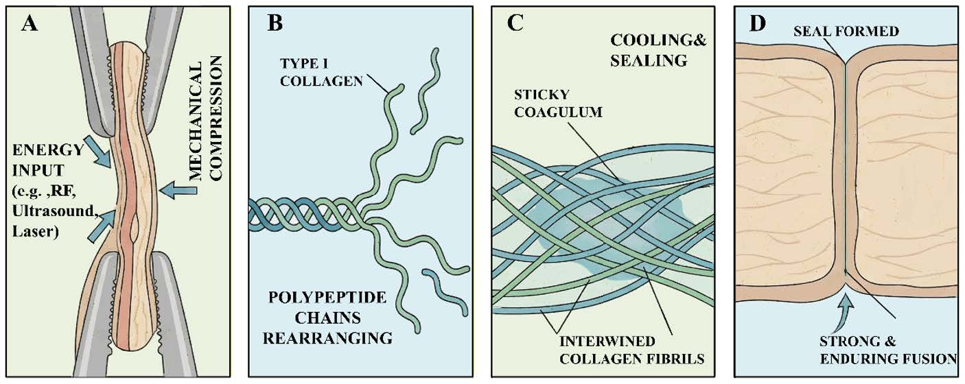
Figure 1. Energy-based tissue fusion mechanism. (A) Energy application and mechanical compression; (B) Collagen denaturation; (C) Cooling and sealing; (D) Tissue fusion. RF, radiofrequency.
Current clinical EBTF technologies predominantly rely on three principal energy modalities: RF energy, ultrasonic energy, and laser energy. Even though EBTF technologies differ a lot as far as how the energy is delivered, they all come together biologically to one common result: using heat to change and rearrange structural proteins in tissues. To get more insight into how each individual modality works, it is essential to first master the mechanisms and evolution of different energy forms.
2.1 RF energy
RF energy refers to alternating electrical currents within the frequency range of 300 kHz to 300 GHz, and is generally regarded as one of the most mature and widely used forms of energy in contemporary electrosurgery. In surgical tissue fusion, current frequency generally varies between 300 kHz and 500 kHz. The surgical use of RF energy primarily revolves around a triad of capabilities: a clean cut, coagulation to stop bleeding, and the sealing of tissues for anastomosis. The high frequency of current notably provides numerous significant benefits: more versatility in use, energy delivery with much higher precision within a shorter period, and a lower probability of neuromuscular stimulation compared with lower-frequency currents.
The core method of RF-driven tissue fusion is based on Joule heating principles. Basically, tissue electrical impedance causes resistance to electrical current flow, which then results in rapid agitation of ions within the cellular and extracellular milieu. The ionic friction thereby directly converts electrical energy into thermal energy, which locally heats the tissue. The effects that consequently occur include both: a non-thermal aspect of electroporation at the level of cell membranes and, more prominently, a deep thermal effect [17]. It is the orderly application of heat that makes the fusion possible.
The history of using RF energy for tissue anastomosis dates back to the early 1990s, when the Paton Welding Institute in Ukraine was the first to conduct pioneering research uniting the concept of tissue welding with controlled energy. In 1998, a major breakthrough came when Kennedy et al. demonstrated, for the first time, the application of RF energy to create an arteriovenous anastomosis, thus proving the possibility of such an approach in vascular tissue [18]. Over the next ten years, the technology went through rapid clinical adoption and development. It was Lamberton et al. who conducted a landmark study that shifted perceptions in 2008, when they compared four laparoscopic vessel ligation devices [19]. Among the devices tested, the RF energy-based system provided the best results in terms of burst pressures and sealing times; therefore, it was confirmed as the most effective for vessel sealing. The technology was also subjected to extensive testing on large animal models. For example, Pan et al. used a porcine model in 2020 to compare RF tissue fusion with traditional suture methods for intestinal anastomosis [20]. Histological examination showed that the gaps in the muscularis layer at the anastomotic site in the RF group were replaced by newly formed collagen fibers. This demonstrates that the tissue was not only sealed but also actively supported during the healing process, thus confirming the method to be a practical and safe alternative.
2.2 Ultrasound
Ultrasonic energy represents another cornerstone modality in EBTF, which is set apart by its special mode of mechanical, rather than electrical, energy conversion. In surgical applications, ultrasonic devices work at very high frequencies, usually between 20 kHz and 60 kHz, well above the limit of human hearing. Contrary to RF energy, which depends on electrical current and heat generated by resistance, ultrasonic emission makes use of piezoelectric transducers in the instrument handle to convert electrical energy into very high-frequency longitudinal mechanical vibrations at the working part of the instrument such as a blade or a jaw [21, 22].
Initially, mechanical friction at a microscopic level is the principal effect when the vibrating blade touches the tissue under the application of a compressive force. Such friction produces a small-scale heat spot exactly at the tissue-blade interface as well as inside the compressed tissue area. The temperature usually rises more slowly and locally in the case of the ultrasonic method than in RF, and the peak temperature is generally only between 80 °C and 100 °C. This thermal effect is a secondary consequence of the primary mechanical energy delivery. The heat causes the denaturation of tissue proteins, mainly collagen and elastin, while the simultaneous, long-lasting pressure and vibration help release water vapor from the tissue. When tissue proteins denature, they create a thick coagulum. The persistent ultrasonic vibration under pressure breaks and reforms the protein bonds, resulting in their realignment and intermixing across the apposed tissue surfaces. They solidify into a single, welded seal on cooling and release of pressure. Very importantly, the significantly lower peak temperatures and the effect of ultrasonic energy in removing water often lead to less lateral thermal damage and less smoke production when compared to RF energy [23].
Ultrasonic energy has shifted from being merely a conceptual instrument to a clinically permitted mode, backed up by an ever-increasing body of research that shows its effectiveness and safety in various surgical fields. In 2002, Foschi et al. used transmission and scanning electron microscopy to study vascular tissue after ultrasonic anastomosis [24]. Their ultrastructural study indicated that the thermal effect caused by ultrasonic energy helps the breakdown and later reformation of tissue proteins, therefore giving a mechanistic explanation of successful tissue fusion. Going from experimentation to clinical testing, Sista et al. performed a comparison study of 211 consecutive patients undergoing right hemicolectomy due to colon cancer [25]. Patients were divided into two groups: one group using a novel ultrasonic energy handpiece (108 patients) and the other group treated with conventional hemostasis methods (103 patients). The data showed that the use of ultrasonic energy notably reduced surgical time, the amount of blood lost during surgery, and postoperative complications. Moreover, Tolone et al. assessed the application of the ultrasonic scalpel for endocrine surgery by comparing total thyroidectomy procedures carried out with an ultrasonic scalpel and conventional suture ligation [26]. Their data demonstrated that ultrasonic energy is a safe, effective, and time-saving alternative. It can facilitate the operations without causing an increase in drainage volume, complications, or length of hospital stay.
2.3 Laser
Laser energy holds a special place in tissue fusion as a separate modality, mainly because of the pure photonic nature of laser. The process of laser generation can be described in simple terms as follows. Electrons in an atomic medium that are excited to a higher energy state by laser light will come down to their ground state and, in doing so, emit photons in a highly synchronized manner. This phenomenon results in the production of light that is monochromatic, coherent, and highly directional [27]. From the surgical point of view, this means that energy can be delivered with extremely high spatial precision, thus the predetermined tissue plane for interaction can be targeted and collateral damage can be minimized. One of the main inherent benefits of laser energy is that, being a form of light, it does not introduce any foreign material to the wound, thus eliminating the main source of infection or chronic inflammatory reactions associated with suture or staple lines.
Laser tissue fusion (LTF) is mainly mediated by the photothermal effect. Thus, when a laser beam at a certain wavelength is absorbed by water or hemoglobin, the energy released is initially converted into heat. This locally generated heat at the spot causes a phase change of the tissue structural proteins. Collagen is one of the proteins in the extracellular matrix that is commonly affected by heat. The triple-helical structure of collagen is mainly maintained by hydrogen bonds. The breaking of these hydrogen bonds occurs at a certain temperature, and this causes the collagen fibrils to lose their structure—going from their highly ordered state into a chaotic, random coil conformation. Importantly, while this process occurs, the tissue’s overall architecture is preserved through mechanical apposition. Following cessation of laser irradiation and during a strict cooling phase, collagen from both tissue surfaces, which is now unfolded and quite mobile, will find each other and, by undergoing recrystallization, form bonds together. Here the polypeptide chains intertwine to form new intermolecular bonds thus producing a uniform, cross-linked protein matrix that closes the wound [28]. It is like a biological “weld” without any gaps, the strength of which comes from the reforged collagen network rather than from the use of foreign materials or mere coagulation necrosis.
The use of laser energy in tissue anastomosis has transitioned through various milestone stages. Firstly, the pioneering work by Jain et al. in 1979 laid down the basis for the feasibility of laser vascular repair [29]. After that, some experiments helped to define the laser welding process parameters more clearly. As an illustration, Bremmer et al. showed that the seal strength depends to a great extent on fine control of laser power, irradiation time, and simultaneous application of pressure in a rabbit artery model [30]. Also, some research articles further detailed the cellular heating mechanisms. For example, Li et al. pointed out that the optimum temperature range for the skin weld is approximately 55–65 °C, above which the mechanical properties become compromised because of significant protein denaturation [31]. Beyond immediate sealing performance, there is still evidence supporting favorable healing outcomes. According to Ghosh et al., laser-sealed mouse skin wounds regained function much earlier than those closed with sutures, suggesting that the laser might support physiological wound healing [32]. All these accomplishments together show the development of LTF from a new idea to a highly technical process. Currently, the emphasis is on broadening its application and integrating in vivo monitoring to achieve better control over the procedure.
The evolution of surgical energy instruments has been one of the paradigm shifts in modern medicine. It signifies a trend from simple anatomical dissection to sophisticated tissue management. As a matter of fact, hemostatic methods over the last 100 years have evolved from suturing and monopolar cautery to the present-day advanced and intelligent tissue fusion platforms. This major change is mostly due to the increasing demand for surgical safety, efficiency, and minimally invasive methods. In this section, we have clinically reviewed several tissue fusion systems that are prevalently used in hospitals and have compared them to each other based on publicly available data from publications indexed in the Science Citation Index Expanded, peer-reviewed biomedical engineering literature, and clinical comparative studies indexed in PubMed (as shown in Table 1). We have focused on explaining the devices’ working principle, preclinical performance metrics, and clinical results in various types of surgery. This multidisciplinary work presents a fair evaluation of the available technologies and points to future innovations required to develop energy-based surgical devices.
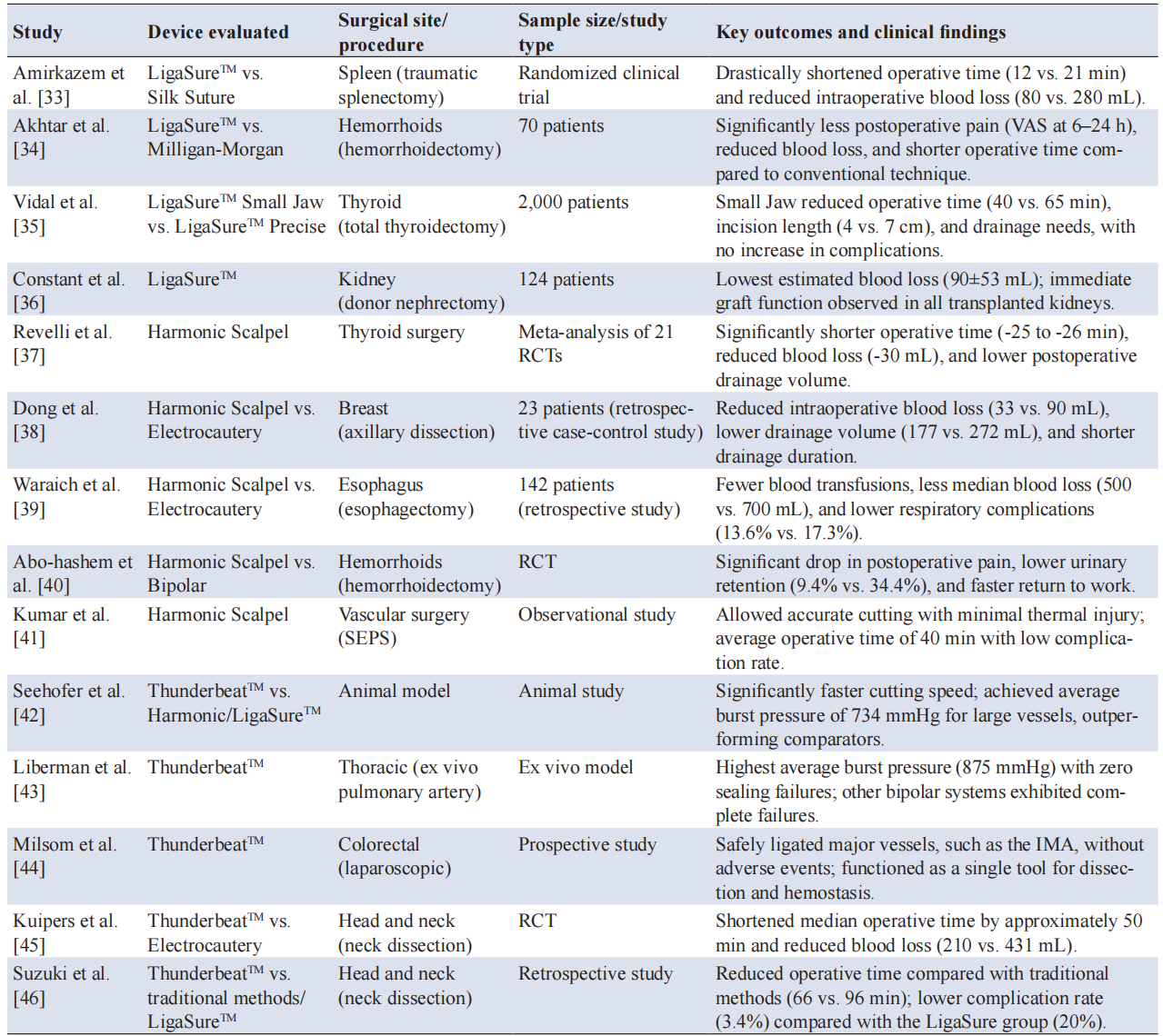
Note: EBTF, energy-based tissue fusion; VAS, visual analog scale; RCT, randomized controlled trial; SEPS, subfascial endoscopic perforator surgery; IMA, inferior mesenteric artery.
3.1 LigaSureTM
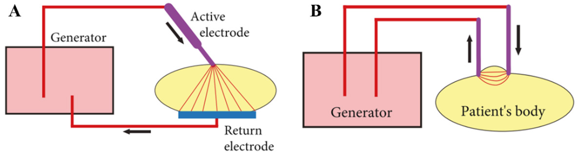
From an engineering viewpoint, the fusion of tissues by means of RF energy is based on two main electrode configurations (as shown in Figure 2). The monopolar configuration uses only one active electrode at the surgical site and the current then flows from the patient’s body to a remote dispersive electrode. This is good for superficial cutting and coagulation, but its wider current path makes it less accurate for tissue fusion. On the other hand, the bipolar configuration has both electrodes incorporated in the instrument jaws. The electric current flows only through the area of the tissue held between them, thus the thermal and electric effects are restricted to a small volume [47]. This concentrated energy release allows the fusion to be very precise, with minimal damage to surrounding tissues, so bipolar systems are suitable for sealing tissues.
To ensure safe surgical seals, many contemporary systems integrate real-time tissue impedance monitoring with automated energy termination. The LigaSureTM system (Medtronic) is a representative example of a closed-loop technology based on tissue impedance monitoring and automated energy termination. Representative LigaSureTM instruments, including the LigaSure ImpactTM and LigaSure AtlasTM, are shown in Figure 3. As a leader in advanced bipolar vessel sealing, LigaSureTM’s main technological feature is its generator platform (ForceTriadTM), which contains the exclusive TissueFectTM sensing system [49]. As technical reports and experimental studies show, the TissueFectTM system measures tissue impedance at a frequency of more than 3,000 measurements per second [50]. The change in tissue impedance induced by the release of intracellular water through vaporization and collagen denaturation follows a typical nonlinear pattern during the sealing cycle. The device continually adjusts voltage and current based on these impedance changes, thus ensuring that the tissue remains within a thermal range that is ideal for fusion while avoiding drying or burning [51]. An algorithm detects an impedance level or changes indicative of seal completion and stops energy delivery automatically. Determining the operative endpoint is fully automated, so subjective surgeon judgment is replaced by objective signal-based control, which in turn leads to improvements in procedural consistency, safety, and seal reliability across different tissue types.
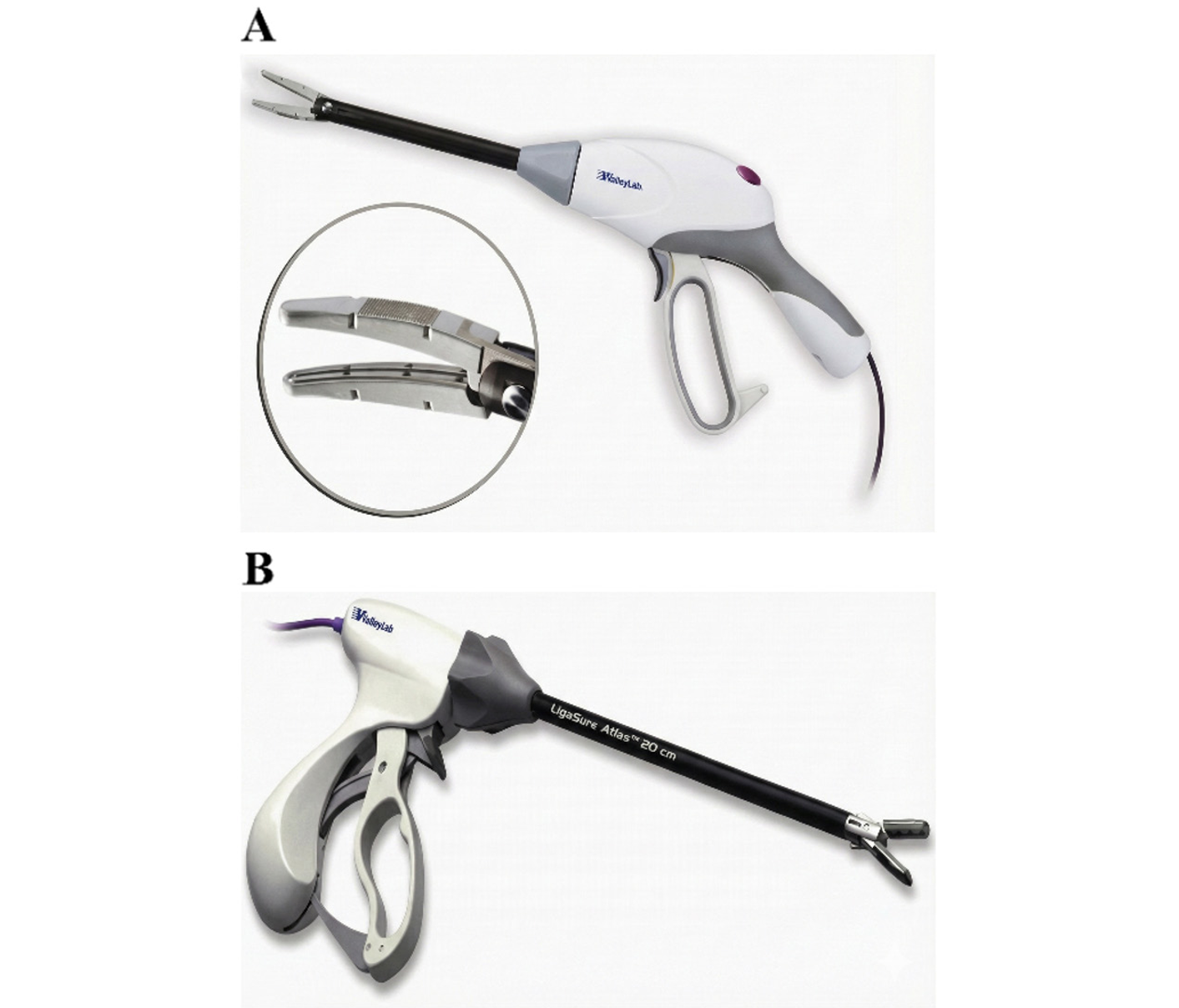
Figure 3. LigaSureTM instruments. (A) LigaSure Impact; (B) LigaSure Atlas. The figure is reproduced from [52].
Numerous randomized controlled trials (RCTs) and retrospective studies within different surgical specialties have confirmed the clinical effectiveness of the LigaSureTM system. Present research highlights three main areas of focus where current evidence is concentrated: more efficient hemostasis superior to traditional methods, device-specific optimization advantages, and safety in complex surgeries.
LigaSureTM, compared with traditional suturing or ligation methods, can significantly shorten operative time and minimize the amount of bleeding during the operation. In a clinical RCT of traumatic splenectomy, the use of LigaSureTM reduced the average duration of the surgery significantly (12 vs. 21 min, P<0.05) and also considerably reduced the intraoperative blood loss (80 vs. 280 mL, P<0.05) as compared with the conventional silk suture ligation [33]. Likewise, in a case of hemorrhoidectomy for grade III/IV hemorrhoids, the device achieved better outcomes than the traditional Milligan-Morgan method as it was not only associated with lower blood loss and shorter operative time but also postoperative pain scores, measured 6–24 hours postoperatively, were significantly lower (P=0.001) [34].
Also, anatomically site-specific models of LigaSureTM have further improved surgical outcomes. According to a retrospective review of 2,000 total thyroidectomy patients, the performance of different generations of devices was compared, and the LigaSureTM Small Jaw—specifically created for use in tight spaces—was found to be better than the standard LigaSure PreciseTM [35]. The Small Jaw was capable of significantly reducing the operative time (40 vs. 65 min, P=0.002), incision size, and the amount of drainage needed without increasing the risk of complications, thus confirming the clinical significance of anatomic device design.
Moreover, in transplant surgery, where the utmost safety precautions are taken, the system has demonstrated reliable vessel sealing functions. In this study of 124 laparoscopic living donor nephrectomies, the device was useful in keeping blood loss to a minimum during the separation of the main vascular branches in the nephrectomy procedure (90±53 mL) [36]. Most importantly, all the transplanted kidneys showed immediate function after reperfusion, thus proving that the device had a good safety record in terms of thermal spread and vessel integrity.
Overall, the research results obtained demonstrate that the LigaSureTM system is a highly versatile and effective advancement in surgical energy devices. It has become an alternative to conventional ligation methods to some extent by continually reducing operative time, blood loss, and postoperative pain, while ensuring high levels of safety in both routine and advanced procedures.
3.2 HARMONICTM
When working in limited anatomical areas or close to vital structures, it is crucial to use surgical energy very precisely. HARMONICTM products are widely recognized as the leader in the ultrasonic energy field (as shown in Figure 4). The device’s principle of operation is radically different from that of bipolar RF devices. Through the piezoelectric effect, a transducer in the handpiece converts electrical energy into mechanical vibration, which causes the blade to oscillate longitudinally at a frequency of 55,500 Hz. Such high-frequency vibration leads to the intensification of molecular friction within tissues. In this way, heat for protein denaturation is produced [49]. Since the heat is produced by internal friction and not by the passage of external electrical current, the device’s temperature is usually lower, and it is significantly lower than that of electrosurgical devices. Moreover, the greatest characteristic of HARMONICTM is that it causes minimal lateral thermal damage. A lot of comparative studies involving both infrared (IR) thermography and histopathology have demonstrated that the lateral thermal spread resulting from the use of Harmonic is usually only about 1–2 mm, which is much less than that of monopolar cautery and some bipolar devices [53]. This feature makes it an excellent tool for delicate dissection in areas with a high concentration of nerves and blood vessels.
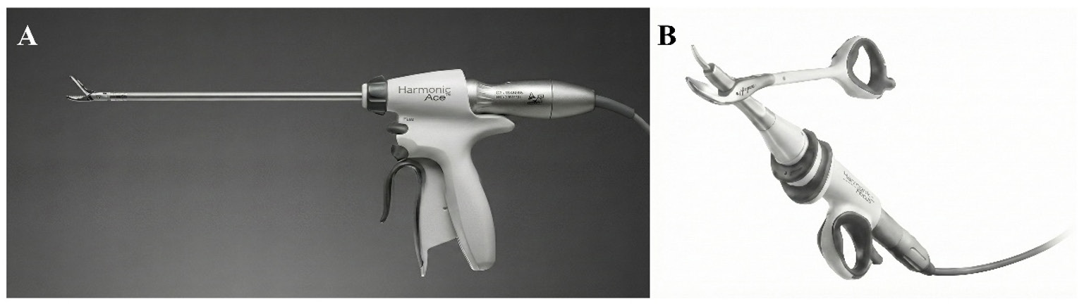
Figure 4. HARMONICTM instruments. (A) Harmonic Ace for laparoscopic use; (B) Harmonic Focus for open use. The figure is reproduced from [54].
It is noteworthy, however, that despite minimal lateral thermal spread, the blade itself can accumulate heat after prolonged activation, necessitating a certain cooling period. Another drawback of the ultrasonic scalpel is that the instrument produces a very visible and substantial surgical smoke (aerosol). Unlike smoke produced by electrocautery, this kind of aerosol consists of larger particle diameters (0.35–6 µm) and has higher levels of moisture and lipid components. Chemical analysis using mass spectrometry (Rapid Evaporative Ionization Mass Spectrometry, REIMS) shows that aerosol is mainly lipids such as diglycerides and triglycerides, as opposed to electrocautery smoke that is rich in glycerophospholipids. Regarding biological risks, even if the chemical toxicity of ultrasonic smoke is lower than that of electrocautery smoke, there is a theoretical risk that cellular materials can be carried because of the larger particle size [55, 56].
The HARMONICTM systems show the traits of versatility and clinical benefits that can be observed across several surgical specialties. The currently available studies point to three major areas of these effects: greater hemostatic effectiveness in oncological and glandular surgeries, an improved post-surgical quality of life, and highly precise vascular interventions.
In particular, during major resections and glandular surgeries, the instrument has been shown to consistently improve fluid management and operative efficiency compared with traditional methods. A meta-analysis of 21 RCTs in thyroid surgery brought out such effectiveness, revealing that the Harmonic Scalpel (HS) and Harmonic Focus notably reduced surgical duration (by 25–26 min, P<0.0001) and reduced blood loss during surgery (by ~30 mL, P<0.01) as compared to conventional hemostasis [37]. These gains have also been supported in breast cancer surgery, where the use of the HS in axillary dissection led to a significantly smaller amount of blood loss during surgery (33 vs. 90 mL, P<0.001) and less drainage volume and duration as compared to electrocautery [38]. Similarly, in esophageal cancer surgery, the device was superior to traditional electrocautery in reducing the need for blood transfusion (median 0 vs. 2 units, P=0.003), reducing blood loss, and lowering the rate of postoperative respiratory complications (13.6% vs. 17.3%) [39].
Moreover, the HS can also significantly improve patient recovery and quality of life long after an operation. In a proctological surgery study, it was found that the HS had markedly better postoperative pain outcomes than bipolar electrocautery [40]. Additionally, it resulted in less frequent use of narcotics and non-steroidal anti-inflammatory drugs, and there was a noticeable decrease in the occurrence of urinary retention (9.4% vs. 34.4%, P<0.05). Therefore, due to earlier discharge and return to work, after only two weeks, 75% of the patients in the HS group had returned to work, compared with 45% in the control group, signifying the socioeconomic benefits of this device.
Furthermore, the fundamental principle of the device’s operation enhances precision in complex vascular interventions. In subfascial endoscopic perforator vein surgery for chronic venous insufficiency, the HS has been demonstrated as an effective alternative to metal clips or electrocautery. By coagulating and cutting simultaneously to produce only a minimal lateral thermal injury, surgeons have been able to achieve precise exposure of perforator veins [41]. The high level of precision here brought about the performance of fast operations (average 40 min) with minimal risk of complications.
In conclusion, HARMONICTM is a unique system that not only improves surgical efficiency but also results in better patient outcomes. The system minimizes tissue harm and controls bleeding which benefits the patient in two ways: on the one hand, it lowers the physiological impact of surgery. As demonstrated by reduced blood loss and drainage in tumor cases. On the other hand, it accelerates patients’ functional recovery, as shown in proctological and vascular procedures.
3.3 ThunderbeatTM
ThunderbeatTM is a hybrid energy system that combines ultrasonic and bipolar RF technologies in one device. Such a design addresses the complementary limitations of each modality: ultrasonic instruments are fast in cutting but have relatively limited hemostasis, whereas bipolar devices, although slower in dissection, provide strong coagulation [49]. As shown in Figure 5, the innovative jaw design of this instrument, consisting of a central ultrasonic probe enclosed by bipolar electrodes, enables the device to operate in two main modes: ‘Seal & Cut’ for simultaneous cutting and sealing, and ‘Seal’ for dedicated coagulation. Preclinical studies in porcine models showed that ThunderbeatTM was able to achieve significantly shorter dissection times than Harmonic Ace, LigaSure V, and EnSeal when working with vessels of various sizes (2–7 mm, P<0.01). Moreover, this device was given a top versatility rating, thus recognizing its ability to streamline surgical procedures through less frequent instrument changes [57].
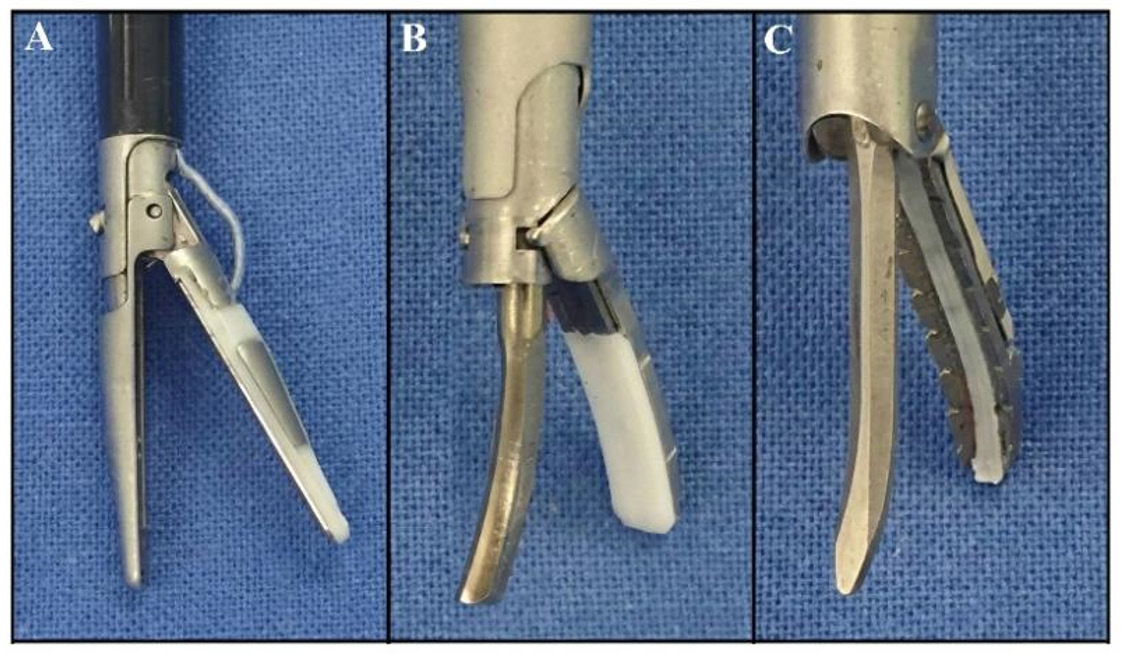
Figure 5. Detailed view of three instruments. (A) LigaSureTM; (B) HARMONICTM; (C) ThunderbeatTM. The figure is reproduced from [42].
The combination of ultrasonic friction and electrical resistance heating, however, accounts for a more intricate thermal footprint. In a direct in vivo porcine model study comparing ThunderbeatTM with EnSeal and LigaSure across multiple tissue types (liver, mesentery, muscle, and spleen), ThunderbeatTM produced significantly higher peak operating temperatures than the other two devices in a consistent manner. It led to the smallest total thermal injury zone in liver tissue, yet its collateral thermal spread in the spleen, mesentery, and muscle was significantly higher than that of EnSeal and LigaSure, which proved to be less thermally intensive and caused negligible collateral damage [58]. Therefore, when dissecting near critical neural or vascular structures, surgeons should take into account the possibility that ThunderbeatTM may have a propensity for greater lateral thermal spread.
There are three key surgical parameters that the existing literature mainly focuses on: the impact of integration on operative speed, the consistency of burst pressure for large vessels, and the efficiency of a single-instrument workflow. Through preclinical studies, the biophysical characteristics of this hybrid technology have been thoroughly defined on a theoretical level, and its dual action of ultrasonic cutting and bipolar coagulation has been demonstrated.
In an animal study, Seehofer et al. found that the device performed cutting operations at a faster rate than single-modality ultrasonic or bipolar devices, yet it was still able to maintain an average burst pressure of 734 mmHg for vessels of 5–7 mm diameter [42]. The sealing capacity was taken to the limit in a model of the lung artery ex vivo by Liberman et al. [43]. Their findings showed that the device was able to close pulmonary branches with an average burst pressure of 875 mmHg, thus demonstrating its potential applicability in the high-pressure vasculature of the chest.
These physical characteristics, when taken together, suggest that the device supports a multipurpose operation workflow, especially during complicated laparoscopic surgeries. Milsom et al. confirmed this aspect of the device in their prospective study on laparoscopic colorectal surgery [44]. They found that the device was able to handle challenging vascular pedicles such as the inferior mesenteric artery and could also be used as the main dissector. Hence, the hybrid instrument concept is supported by this case where the use of different instruments can be avoided by combining dissection and hemostasis into one single operation.
Versatility of these devices translates most advantageously to head and neck surgical settings in terms of documented operative time. In this domain, the highest level of evidence exists. Kuipers et al., comparing the device with traditional electrocautery, measured a median reduction in operative time of almost 50 minutes besides a decrease in blood loss (210 mL vs. 431 mL) [45]. To add to that, Suzuki et al. in a retrospective study looked at the device versus two other methods: the traditional approach and the LigaSureTM system [46]. While noticing shorter surgery duration as well, this research pinpointed the fact that the ThunderbeatTM cohort encountered fewer complications (3.4%) than the LigaSureTM group (20%). Taken together, these results imply that the main clinical benefit of the device is the reduction of operative time without compromising safety.
In essence, the ThunderbeatTM system is an effective alternative in the energy device market. Marrying ultrasonic speed with bipolar hemostasis, it delivers a device that is not only highly efficient but also very flexible, thus meeting a wide range of surgical situations such as, for example, procedures that require both thorough dissection and safe handling of bigger blood vessels, thereby assisting in attaining surgical efficiency in different disciplines.
3.4 Laser-based tissue fusion systems
Unlike RF and ultrasonic devices that rely on mechanically compressive jawed instruments, LTF or laser tissue soldering (LTS) primarily utilizes non-contact fiber-optic delivery systems combined with precise optical and thermal sensors. The fundamental engineering challenge in laser-based equipment is temperature regulation. The success of laser fusion depends entirely on maintaining the tissue surface temperature within a very narrow therapeutic window (typically 60 °C to 65 °C for optimal collagen denaturation). Overheating causes irreversible thermal necrosis and tissue vaporization, while underheating leads to insufficient protein cross-linking and weak tissue bonds.
To address this, modern clinical laser fusion equipment heavily relies on closed-loop temperature control systems. A typical temperature-controlled laser soldering system incorporates a laser generator (e.g., carbon dioxide, neodymium-doped yttrium aluminum garnet, or near-IR diode lasers), a flexible delivery optical fiber, and a real-time IR thermometry circuit or thermal imaging feedback circuit [59]. During the procedure, the IR sensor continuously monitors the thermal footprint at the tissue interface. A control algorithm processes this data in real-time to dynamically modulate the laser power output, ensuring that the tissue temperature stays within the target range without manual intervention.
Such systems have made a smooth transition to clinical applications. For example, during a pilot clinical trial of laparoscopic cholecystectomy, the temperature-controlled laser soldering system was used to close human skin incisions. The system was regularly successful in producing sealed wounds. Besides, the net time for closure was significantly reduced compared with traditional suturing methods, thus confirming the clinical feasibility of temperature-controlled laser devices [60].
Furthermore, the latest frontier in laser fusion equipment involves the integration of these temperature-controlled optical systems with surgical robotic platforms, known as robot-assisted LTS systems [61]. By mounting the laser applicator and IR sensors onto robotic arms, robot-assisted LTS systems eliminate the inconsistencies caused by human hand tremors and variable scanning speeds. This automated, high-precision control over both the spatial delivery of laser energy and the thermal parameters yields highly reproducible tissue bonds, paving the way for advanced, semi-automated wound closure in minimally invasive surgeries.
As EBTF technologies mature, the next frontier in surgical innovation will be the transition from passive energy delivery tools to active, intelligent surgical platforms. This shift is enabled by perfectly combining AI, machine learning, and advanced robotic systems.
4.1 AI-driven energy modulation and real-time tissue identification
At present, the majority of EBTF devices use simple impedance feedback to stop energy delivery. Nevertheless, the use of machine learning algorithms can bring about highly dynamic energy modulation. Through the constant analysis of multi-modal sensor inputs, e.g., tissue impedance, thermal dissipation, and hydration levels, AI models are able to forecast the best sealing endpoint for heterogeneous tissues instantly, thus achieving maximum hemostasis while minimizing iatrogenic thermal injury.
Furthermore, a groundbreaking application of AI in EBTF is the real-time identification of tissues through the analysis of surgical smoke. When EBTF devices cut or coagulate tissue, they generate aerosols rich in cellular metabolites and lipids. Technologies such as REIMS—often referred to as the “iKnife” concept—can capture this smoke. By coupling REIMS with machine learning classifiers (such as principal component an-
alysis and neural networks), the system can analyze the mass spectrometric profile in milliseconds [62]. This allows the EBTF instrument to act as a smart diagnostic probe, instantly distinguishing between healthy tissue, critical structures (like nerves and blood vessels), and malignant tumor margins during oncological surgeries.
4.2 Integration with robotic surgical systems
The physical execution of tissue fusion is also being revolutionized by robotic platforms, such as the da Vinci Surgical System. Integrating advanced EBTF instruments—like fully articulated robotic vessel sealers—into robotic arms overcomes the physical limitations of human surgeons. Robotic integration offers three-dimensional high-definition visualization, eliminates physiological hand tremors, and provides a 360-degree range of motion. This allows for unparalleled precision when applying energy and mechanical compression in confined, complex anatomical spaces, such as the deep pelvis or the pancreatic head [63].
Looking ahead, the fusion of AI-based computer vision and robotic EBTF devices will allow for semi-autonomous surgical maneuvers. It is anticipated that future robotic systems will be able to locate vessels automatically, measure the precise pressure needed, and deliver the specific energy dosage for each patient, thereby ensuring consistent surgical results and lowering the mental burden on surgeons.
In short, tissue closure technology has seen a drastic change from traditional mechanical anastomosis to EBTF, which is nowadays regarded as a significant paradigm shift in surgical practice. The article has deeply discussed the mechanism of EBTF, which uses controlled thermal energy to cause collagen denaturation and molecular rearrangement, thus avoiding the major problems of sutures and staplers still commonly used today, such as foreign body reactions and technical difficulties in minimally invasive surgeries. It does not really matter if the tissue is heated through Joule heating by RF energy, through the mechanical friction of ultrasonic vibrations, or through the photothermal effect of laser energy; biologically, all these methods target the same outcome: a strong, self-healing tissue seal. Taken to clinical practice, these concepts have led to the development of revolutionary medical devices. A good example of this is the LigaSureTM system which uses a real-time impedance feedback technology to continuously and accurately seal tissues. The HARMONICTM system relies on ultrasonic vibrations to precisely cut tissue while effectively limiting lateral thermal damage. The ThunderbeatTM system is a hybrid of the two techniques that offers the speed of ultrasound combined with the safety of bipolar sealing. The large amount of clinical data presented in this paper demonstrates that these techniques are not only effective in achieving hemostasis but also, through their associated medical devices, greatly facilitate the surgeon’s work, reduce the duration of the operation, and minimize blood loss. On top of that, they have such a wide range of application in basically every specialty that they can be used in procedures ranging from simple vessel ligation to digestive, endocrine, and urological surgeries.
Looking forward, the development of energy-based surgical devices will be focused on more than just the power generation aspect. These devices will be more intelligent, precise, and integrated with robotic technologies [64]. Energy devices will become seamlessly integrated with robotic systems as the use of robotic platforms for minimally invasive surgery increases. Although advanced energy-based devices are associated with higher costs at the initial stage, their capability of lessening the duration of the operation and the rate of complications is translated into a favorable long-term cost-benefit scenario for the healthcare system [65]. Furthermore, the use of AI and computer vision is expected to completely change the concept of surgical safety. Intelligent surgical systems of the future will most probably use machine learning algorithms to identify the nature of tissues during surgery in real-time [66]. As a result, next-generation surgical devices might be capable of automatically identifying nerves, blood vessels, or other tissues. In this way, they can dynamically adjust energy output to maximize hemostasis while minimizing iatrogenic thermal injury. Ultimately, these innovations are intended to be able to strike the perfect combination of anastomotic strength, minimal tissue injury, and optimal healing results.
Author contributions
Junjie Shen and Zhongxin Hu were responsible for writing the original draft and conducting the investigation. Lin Mao and Chengli Song were responsible for writing, reviewing, and editing the manuscript, as well as supervising the project.
Funding
This research received no external funding.
Data availability
No new data were generated or analyzed in this review. All data cited in this article are from previously published studies, which have been appropriately referenced in the text.
Ethics approval and consent to participate
Not applicable.
Consent for publication
All authors acknowledge and agree to submit this article to the journal Progress in Medical Devices.
Competing interests
The authors declare that they have no competing interests.
Acknowledgements
Not applicable.
[1] Banga A, Saeed Muhammad S, Misra S, Ying X, Patino C, Rathore Sawai S, et al. Sealing the deal: A meta-analysis of suture-only and hybrid closure techniques for femoral access hemostasis after percutaneous cardiovascular procedures. J Am Coll Cardiol. 2025 Apr 1;85(12_Supplement):854-854. https://doi.org/10.1016/S0735-1097(25)01338-5
[2] Chen Y, Chai M, Xuan C, Lin J, Yang H, Li C, et al. Tuning the properties of surgical polymeric materials for improved soft-tissue wound closure and healing. Prog Mater Sci. 2024 Jun 1;143:101249. https://doi.org/10.1016/j.pmatsci.2024.101249
[3] Crumplin MKH. Surgical suture needles. Br J Surg. 2023 Mar 2;110(5):548-552. https://doi.org/10.1093/bjs/znad053
[4] Yang N, Venezuela J, Allavena R, Lau C, Dargusch M. Zinc-based subcuticular absorbable staples: An in vivo and in vitro study. Acta Biomater. 2023 Sep 1;167:593-607. https://doi.org/10.1016/j.actbio.2023.06.030
[5] Albeniz E, Alvarez MA, Espinos JC, Nogales O, Guarner C, Alonso P, et al. Clip closure after resection of large colorectal lesions with substantial risk of bleeding. Gastroenterology. 2019 Nov 1;157(5):1213-1221. https://doi.org/10.1053/j.gastro.2019.07.037
[6] Pagán AJ, Ramakrishnan L. The formation and function of granulomas. Annu Rev Immunol. 2018 Feb 5;36:639-665. https://doi.org/10.1146/annurev-immunol-032712-100022
[7] Gong Y, Li C, Zhu B, Li F, Feng L, Zhao Y, et al. Engineering injectable bioadhesives with sealing and anti-fouling properties for postoperative anti-adhesion. Chem Eng J. 2024 Nov 15;500:156747. https://doi.org/10.1016/j.cej.2024.156747
[8] Kelmansky R, McAlvin BJ, Nyska A, Dohlman JC, Chiang HH, Hashimoto M, et al. Strong tissue glue with tunable elasticity. Acta Biomater. 2017 Apr 15;53:93-99. https://doi.org/10.1016/j.actbio.2017.02.009
[9] Peng W, Lai Y, Fan J, Dai C, Shen J, Liu P. Simple component self-gelling powders as multifunctional tissue adhesives for sutureless wound healing. Chem Eng J. 2024 Sep 1;495:153514. https://doi.org/10.1016/j.cej.2024.153514
[10] Zheng Y, Baidya A, Annabi N. Molecular design of an ultrastrong tissue adhesive hydrogel with tunable multifunctionality. Bioact Mater. 2023 Nov 1;29:214-229. https://doi.org/10.1016/j.bioactmat.2023.06.007
[11] Xu F, Lu T, Seffen KA. Biothermomechanical behavior of skin tissue. Acta Mech Sin. 2008 Feb 1;24(1):1-23. https://doi.org/10.1007/s10409-007-0128-8
[12] Leo L, Bridelli MG, Polverini E. Reversible processes in collagen dehydration: A molecular dynamics study. Arch Biochem Biophys. 2021 Dec 15;714:109079. https://doi.org/10.1016/j.abb.2021.109079
[13] Mogilner IG, Ruderman G, Grigera JR. Collagen stability, hydration and native state. J Mol Graph Model. 2002 Dec 1;21(3):209-213. https://doi.org/10.1016/S1093-3263(02)00145-6
[14] Wright NT, Humphrey JD. Denaturation of collagen via heating: An irreversible rate process. Annu Rev Biomed Eng. 2002 Jul 16;4:109-128. https://doi.org/10.1146/annurev.bioeng.4.101001.131546
[15] Kramer EA, Rentschler ME. Energy-based tissue fusion for sutureless closure: Applications, mechanisms, and potential for functional recovery. Annu Rev Biomed Eng. 2018 Jun 4;20:1-20. https://doi.org/10.1146/annurev-bioeng-071516-044702
[16] Su L, Cloyd KL, Arya S, Hedegaard MA, Steele JA, Elson DS, et al. Raman spectroscopic evidence of tissue restructuring in heat-induced tissue fusion. J Biophotonics. 2013 Nov 15;7(9):713-723. https://doi.org/10.1002/jbio.201300099
[17] Zhou D, Yang G, Xu J, Ling B, Wang S. Non-thermal effect of radio frequency treatments verified by the multi-scale structure and in-vitro digestibility of sweet potato starch. Innov Food Sci Emerg Technol. 2023 Jul 1;87:103412. https://doi.org/10.1016/j.ifset.2023.103412
[18] Kennedy JS, Stranahan PL, Taylor KD, Chandler JG. High-burst-strength, feedback-controlled bipolar vessel sealing. Surg Endosc. 1998 Jun 1;12(6):876-878. https://doi.org/10.1007/s004649900733
[19] Lamberton GR, Hsi RS, Jin DH, Lindler TU, Jellison FC, Baldwin DD. Prospective comparison of four laparoscopic vessel ligation devices. J Endourol. 2008 Oct 1;22(10):2307-2312. https://doi.org/10.1089/end.2008.9715
[20] Pan H, Leung KKC, Ng EKW. Tissue fusion technology versus suture and staple in porcine bowel anastomosis: An in vivo study. Braz J Med Biol Res. 2020 Apr 17;53(5):e9305. https://doi.org/10.1590/1414-431X20209305
[21] Cardoso GS, Martins YA, Lopez RFV, Carneiro AAO, Lopes JH, Pavan TZ. Medical and biological applications of Langevin-type ultrasonic transducers: A narrative review. Ultrasonics. 2026 Feb 1;158:107825. https://doi.org/10.1016/j.ultras.2025.107825
[22] Saber Jahromi M, Abedini R. Design optimization and performance characteristics of a hemostatic ultrasonic scalpel. Int J Interact Des Manuf. 2025 Sep 1;19(9):6319-6330. https://doi.org/10.1007/s12008-024-02216-z
[23] Yang X, Cao J, Yan Y, Liu F, Li T, Han L, et al. Comparison of the safety of electrotome, Harmonic scalpel, and LigaSure for management of thyroid surgery. Head Neck. 2017 Jun 1;39(6):1078-1085. https://doi.org/10.1002/hed.24701
[24] Foschi D, Cellerino P, Corsi F, Taidelli T, Morandi E, Rizzi A, et al. The mechanisms of blood vessel closure in humans by the application of ultrasonic energy. Surg Endosc Other Interv Tech. 2002 May 1;16(5):814-819. https://doi.org/10.1007/s00464-001-9074-x
[25] Sista F, Abruzzese V, Schietroma M, Cecilia EM, Mattei A, Amicucci G. New harmonic scalpel versus conventional hemostasis in right colon surgery: A prospective randomized controlled clinical trial. Dig Surg. 2013 Jan 1;30(4-6):355-361. https://doi.org/10.1159/000354864
[26] Tolone S, Bondanese M, Ruggiero R, Gili S, Pirozzi R, Parisi S, et al. Outcomes of sutureless total thyroidectomy in elderly. Int J Surg. 2016 Sep 1;33 Suppl 1:S16-S19. https://doi.org/10.1016/j.ijsu.2016.05.056
[27] Wright VC, Riopelle MA. Laser physics for surgeons: An introduction to carbon dioxide laser surgery. Acta Obstet Gynecol Scand. 1984 Jan 1;63(S125):5-15. https://doi.org/10.3109/00016348409157012
[28] Bass LS, Moazami N, Pocsidio J, Oz MC, Logerfo P, Treat MR. Changes in type I collagen following laser welding. Lasers Surg Med. 1992 Jan 1;12(5):500-505. https://doi.org/10.1002/lsm.1900120508
[29] Jain KK, Gorisch W. Repair of small blood vessels with the neodymium-YAG laser: A preliminary report. Surgery. 1979 Jun;85(6):684-688.
[30] Bremmer J, van Doormaal TPC, Verweij BH, van der Zwan A, Tulleken CAF, Verdaasdonk R. Vessel wall perforation mechanism of the excimer laser-assisted non-occlusive anastomosis technique. Lasers Med Sci. 2016 May 25;31(6):1169-1175. https://doi.org/10.1007/s10103-016-1950-7
[31] Li C, Wang K. Effect of welding temperature and protein denaturation on strength of laser biological tissues welding. Opt Laser Technol. 2021 Jun 1;138:106862. https://doi.org/10.1016/j.optlastec.2020.106862
[32] Ghosh D, Salinas CM, Pallod S, Roberts J, Makin IRS, Yaron JR, et al. Temporal evaluation of efficacy and quality of tissue repair upon laser-activated sealing. Bioeng Transl Med. 2022 Sep 28;8(2):e10412. https://doi.org/10.1002/btm2.10412
[33] Amirkazem VS, Malihe K. Randomized clinical trial of ligasure™ versus conventional splenectomy for injured spleen in blunt abdominal trauma. Int J Surg. 2017 Feb 1;38:48-51. https://doi.org/10.1016/j.ijsu.2016.12.036
[34] Akhtar F, Rauf S, Noor A, Ayubi A, Sakhizada F. 314 Comparison of hemorrhoidectomy by LigaSure and conventional Milligan Morgan's technique. Br J Surg. 2023 Aug 21;110(Supplement n.6):znad241.478. https://doi.org/10.1093/bjs/znad241.478
[35] Vidal O, Saavedra-Perez D, Valentini M, Astudillo E, Fernández-Cruz L, Garcia-Valdecasas JC. Surgical outcomes of total thyroidectomy using the LigaSure™ Small Jaw versus LigaSure PreciseTM: A retrospective study of 2000 consecutive patients. Int J Surg. 2017 Jan 1;37:8-12. https://doi.org/10.1016/j.ijsu.2016.08.241
[36] Constant DL, Florman SS, Mendez F, Thomas R, Slakey DP. Use of the LigaSure vessel sealing device in laparoscopic living-donor nephrectomy. Transplantation. 2004 Dec 15;78(11):1661-1664. https://doi.org/10.1097/01.tp.0000144379.29943.ed
[37] Revelli L, Damiani G, Bianchi CBNA, Vanella S, Ricciardi W, Raffaelli M, et al. Complications in thyroid surgery. Harmonic scalpel, harmonic focus versus conventional hemostasis: A meta-analysis. Int J Surg. 2016 Apr 1;28:S22-S32. https://doi.org/10.1016/j.ijsu.2015.12.050
[38] Dong W. 87P Therapeutic effect of harmonic scalpel versus electrocautery in axillary dissection of breast cancer. Ann Oncol. 2016 Dec 1;27:25-26. https://doi.org/10.1016/S0923-7534(21)00247-7
[39] Waraich N, Ahmed J, Rashid F, Mulvey D, Leeder P, Iftikhar SY. Is harmonic scalpel an effective tool for oesophagectomy? Int J Surg. 2009 Jan 1;7(4):330-333. https://doi.org/10.1016/j.ijsu.2009.03.001
[40] Abo-hashem AA, Sarhan A, Aly AM. Harmonic Scalpel® compared with bipolar electro-cautery hemorrhoidectomy: A randomized controlled trial. Int J Surg. 2010 Jan 1;8(3):243-247. https://doi.org/10.1016/j.ijsu.2010.01.010
[41] Kumar A, Agarwal PN, Kumar Garg P. Evaluation of subfascial endoscopic perforator vein surgery (SEPS) using harmonic scalpel in varicose veins: An observational study. Int J Surg. 2009 Jan 1;7(3):253-256. https://doi.org/10.1016/j.ijsu.2009.04.005
[42] Seehofer D, Mogl M, Boas-Knoop S, Unger J, Schirmeier A, Chopra S, et al. Safety and efficacy of new integrated bipolar and ultrasonic scissors compared to conventional laparoscopic 5-mm sealing and cutting instruments. Surg Endosc. 2012 Mar 27;26(9):2541-2549. https://doi.org/10.1007/s00464-012-2229-0
[43] Liberman M, Khereba M, Goudie E, Kazakov J, Thiffault V, Lafontaine E, et al. Pilot study of pulmonary arterial branch sealing using energy devices in an ex vivo model. J Thorac Cardiovasc Surg. 2014 Aug 16;148(6):3219-3223. https://doi.org/10.1016/j.jtcvs.2014.05.089
[44] Milsom JW, Trencheva K, Sonoda T, Nandakumar G, Shukla PJ, Lee S. A prospective trial evaluating the clinical performance of a novel surgical energy device in laparoscopic colon surgery. Surg Endosc. 2015 Aug 27;29(5):1161-1166. https://doi.org/10.1007/s00464-014-3783-4
[45] Kuipers NC, de Kleijn BJ, Wedman J, van der Laan B, Plaat BEC, Halmos GB. A comparison of the Thunderbeat and standard electrocautery devices in head and neck surgery: A prospective randomized controlled trial. Eur Arch Otorhinolaryngol. 2021 Mar 19;278(12):4987-4996. https://doi.org/10.1007/s00405-021-06739-z
[46] Suzuki K, Shimizu M, Sakagami T, Yagi M, Fujisawa T, Sawada S, et al. A comparison of short-term outcomes of neck dissection for head and neck cancers using Thunderbeat, LigaSure or treatment without an energy-based device: A case controlled study. Int J Surg. 2018 Sep 21;58:60-64. https://doi.org/10.1016/j.ijsu.2018.09.009
[47] Marynskyy GS, Tkachenko VA, Bysko VO, Podpryatov SE, Podpritatov SS, Grabovskyi SD, et al. High-frequency equipment for live tissue welding. Paton Weld J. 2023 Jan 4;1(1):23-30. https://doi.org/10.37434/tpwj2023.01.04
[48] Taheri A, Mansoori P, Sandoval LF, Feldman SR, Pearce D, Williford PM. Electrosurgery: part II. Technology, applications, and safety of electrosurgical devices. J Am Acad Dermatol. 2014 Mar 13;70(4):607.e1-607.e12. https://doi.org/10.1016/j.jaad.2013.09.055
[49] Sran H, Sebastian J, Hossain MA. Electrosurgical devices: Are we closer to finding the ideal appliance? A critical review of current evidence for the use of electrosurgical devices in general surgery. Expert Rev Med Devices. 2016 Feb 1;13(2):203-215. https://doi.org/10.1586/17434440.2016.1134312
[50] Sutton PA, Awad S, Perkins AC, Lobo DN. Comparison of lateral thermal spread using monopolar and bipolar diathermy, the Harmonic ScalpelTM and the LigasureTM. Br J Surg. 2010 Jan 27;97(3):428-433. https://doi.org/10.1002/bjs.6901
[51] Palazzo FF, Francis DL, Clifton MA. Randomized clinical trial of LigasureTM versus open haemorrhoidectomy. Br J Surg. 2002 Feb 22;89(2):154-157. https://doi.org/10.1046/j.1365-2168.2002.01993.x
[52] Schuchert MJ, Abbas G, Landreneau JP, Luketich JD, Landreneau RJ. Use of energy-based coagulative fusion technology and lung sealants during anatomic lung resection. J Thorac Cardiovasc Surg. 2012 Sep 1;144(3):S48-S51. https://doi.org/10.1016/j.jtcvs.2012.06.001
[53] Manoli E, Mason S, Ford L, Adebesin A, Bodai Z, Darzi A, et al. Validation of ultrasonic harmonic scalpel for real-time tissue identification using rapid evaporative ionization mass spectrometry. Anal Chem. 2021 Apr 13;93(14):5906-5916. https://doi.org/10.1021/acs.analchem.1c00270
[54] Nanashima A, Abo T, Arai J, Takagi K, Matsumoto H, Takeshita H, et al. Usefulness of vessel-sealing devices combined with crush clamping method for hepatectomy: A retrospective cohort study. Int J Surg. 2013 Nov 1;11(9):891-897. https://doi.org/10.1016/j.ijsu.2013.07.012
[55] Krones CJ, Conze J, Hoelzl F, Stumpf M, Klinge U, Möller M, et al. Chemical composition of surgical smoke produced by electrocautery, harmonic scalpel and argon beaming - a short study. Eur Surg. 2007 Apr 1;39(2):118-121. https://doi.org/10.1007/s10353-006-0305-1
[56] Barrett WL, Garber SM. Surgical smoke: A review of the literature. Surg Endosc Other Interv Tech. 2003 Jun 1;17(6):979-987. https://doi.org/10.1007/s00464-002-8584-5
[57] Milsom J, Trencheva K, Monette S, Pavoor R, Shukla P, Ma J, et al. Evaluation of the safety, efficacy, and versatility of a new surgical energy device (THUNDERBEAT) in comparison with Harmonic ACE, LigaSure V, and EnSeal devices in a porcine model. J Laparoendosc Adv Surg Tech. 2012 May 1;22(4):378-386. https://doi.org/10.1089/lap.2011.0420
[58] Dunay MP, Lipcsey Z, Arany-Tóth A, Nemeth T, Solymosi N, Venczel L, et al. Experimental assessment of three electrosurgical tissue-sealing devices in a porcine model. Acta Vet Hung. 2020 Sep 30;68(3):318-322. https://doi.org/10.1556/004.2020.00051
[59] Dizman B, Ruhi MK. Development of an infrared array sensor-integrated laser system for precision and efficacy in medical applications. Lasers Med Sci. 2025 Jun 5;40(1):254. https://doi.org/10.1007/s10103-025-04510-y
[60] David Simhon MD, Ilan G, Gregory S, Tamar V, Israel N, Roberto M, et al. Temperature-controlled laser-soldering system and its clinical application for bonding skin incisions. J Biomed Opt. 2015 Dec 1;20(12):128002. https://doi.org/10.1117/1.JBO.20.12.128002
[61] Basov S, Milstein A, Sulimani E, Platkov M, Peretz E, Rattunde M, et al. Robot-assisted laser tissue soldering system. Biomed Opt Express. 2018 Nov 1;9(11):5635-5644. https://doi.org/10.1364/BOE.9.005635
[62] Balog J, Sasi-Szabó L, Kinross J, Lewis MR, Muirhead LJ, Veselkov K, et al. Intraoperative tissue identification using rapid evaporative ionization mass spectrometry. Sci Transl Med. 2013 Jul 17;5(194):194ra193. https://doi.org/10.1126/scitranslmed.3005623
[63] Abaza R, Henderson SJ, Martinez O. Robotic vessel sealer device for lymphocele prevention after pelvic lymphadenectomy: Results of a randomized trial. J Laparoendosc Adv Surg Tech. 2022 May 31;32(7):721-726. https://doi.org/10.1089/lap.2021.0531
[64] Saeidi H, Opfermann JD, Kam M, Wei S, Leonard S, Hsieh MH, et al. Autonomous robotic laparoscopic surgery for intestinal anastomosis. Sci Robot. 2022 Jan 26;7(62):eabj2908. https://doi.org/10.1126/scirobotics.abj2908
[65] Vettorettto N, Foglia E, Gerardi C, Lettieri E, Nocco U, Botteri E, et al. High-energy devices in different surgical settings: Lessons learnt from a full health technology assessment report developed by SICE (Società Italiana di Chirurgia Endoscopica). Surg Endosc. 2023 Apr 1;37(4):2548-2565. https://doi.org/10.1007/s00464-022-09734-5
[66] Hashimoto DA, Rosman G, Rus D, Meireles OR. Artificial intelligence in surgery: Promises and perils. Ann Surg. 2018 Jul 1;268(1):70-76. https://doi.org/10.1097/sla.0000000000002693
ISSN: 2957-5478
Volume 4, Issue 2
June 2026
Pages: 77-147


 Log In

 On This Page
 CITE
CITE