 Log In
Search




 Open Access
 yanshj99@aliyun.com
Shiju Yan, School of Health Science and Engineering, University of Shanghai for Science and Technology, No. 334 Jungong Road, Yangpu District, Shanghai 200093, China. E-mail: yanshj99@aliyun.com.
Received December 3, 2025; Accepted April 30, 2026; Published June 18, 2026
Open Accessyanshj99@aliyun.comShiju Yan, School of Health Science and Engineering, University of Shanghai for Science and Technology, No. 334 Jungong Road, Yangpu District, Shanghai 200093, China. E-mail: yanshj99@aliyun.com.
Received December 3, 2025; Accepted April 30, 2026; Published June 18, 2026
Endoscopic submucosal dissection (ESD) is now considered the standard endoscopic resection technique for patients with early gastric cancer. ESD provides a higher rate of complete resection and a lower local recurrence rate. However, ESD results in larger and deeper ulcers, and post-ESD bleeding is a common complication. Bleeding after ESD cannot be completely avoided, especially in patients with large-sized gastric ulcers, those on anticoagulant therapy, and elderly patients. Most bleeding can be controlled by endoscopic hemostatic methods during the procedure, such as the application of hemostatic clips for ulcer closure and hemostatic powder for ulcer shielding. In addition, we also found the potential value of using new materials such as self-assembling peptides for hemostasis. This review first revisits the definition of endoscopic resection of the digestive tract. Then, we discuss post-ESD bleeding and the influence of risk factors such as the location, size, and depth of the surgical lesion, anticoagulant medication use, and the patient's age and lifestyle. Finally, we review the treatment methods for post-ESD bleeding, including intraoperative ulcer closure, ulcer shielding, and the application of thrombin and adrenaline injection.
Keywords: Digestive tract tumors, Digestive tract endoscopy, Bleeding risk factors, Hemostasis in gastric endoscopic submucosal dissection
Esophageal cancer and gastric cancer are common malignant tumors of the digestive system. Patients who can be diagnosed and treated in the early stage generally have a relatively good prognosis. Atypical hyperplasia of the esophageal and gastric mucosa is a precancerous lesion of esophageal cancer and gastric cancer. Early diagnosis and treatment of atypical hyperplasia of the esophageal and gastric mucosa can block the development of precancerous lesions into esophageal cancer and gastric cancer.
Endoscopic resection is now widely accepted as a less invasive treatment for local resection of early gastric cancer, with a negligible risk of lymph node metastasis [1]. A suitable endoscopic resection technique should be safe, effective, and applicable to various clinical situations. However, endoscopic resection is associated with various complications, the most important of which are bleeding and perforation [2].
Endoscopic resection for digestive tract lesions has the advantages of minimal surgical trauma, fewer postoperative complications, and good postoperative recovery, making it the preferred minimally invasive surgical treatment for digestive tract mucosal and submucosal lesions [3].
Endoscopic mucosal resection (EMR) and endoscopic submucosal dissection (ESD) are two endoscopic resection procedures for gastrointestinal lesions. Compared with standard polypectomy techniques, EMR and ESD provide wider and deeper resection margins and allow for more detailed pathological evaluation of the overall resected lesion, which has advantages in treating early neoplastic gastrointestinal lesions [3]. Although EMR is convenient, larger lesions cannot be completely resected in a single EMR procedure. Therefore, doctors cannot make an accurate diagnosis of the entire pathological specimen to make more appropriate treatment decisions for the patient’s condition, which increases the risk of local tumor recurrence or inappropriate treatment. ESD can directly resect tumors in the submucosa. Due to the large wound size and the difficulty of implementing the technique, there is a higher incidence of bleeding and perforation after ESD [4].
This review focuses on post-ESD bleeding by summarizing relevant literature on ESD, including surgical procedures, risk factors for delayed bleeding, prevention and treatment strategies, and related medical devices.
2.1 Definitions and pathophysiology
In this review, the literature search was carried out through the Web of Science database, and the search time range was set from the establishment of the database to September 2025. The core search terms included “Endoscopic Submucosal Dissection”, “Gastrointestinal Hemorrhage”, “Immediate Bleeding”, “Delayed Bleeding”, and “Risk Factors”. According to the onset time, bleeding complications can be subdivided into immediate (intraoperative) bleeding that occurs during the procedure and delayed bleeding that occurs after the procedure. There are differences in risk factors and treatment logic between these two types of hemorrhage. Immediate hemorrhage is mainly affected by anatomical location and procedural factors, while delayed hemorrhage is more related to the patient’s general condition and healing environment. Immediate bleeding is infrequent with EMR techniques, but is quite common with ESD. The management of immediate bleeding plays a key role in the successful completion of ESD. It is difficult to accurately measure the amount of bleeding during EMR and ESD. Therefore, in the definitions of upper gastrointestinal bleeding (GIB) in different countries, bleeding after ESD is usually defined as a condition with any clinical signs of bleeding. Clinical signs include hematemesis, melena, or a decrease in haemoglobin of >2 g/dL from the previous laboratory test, and the condition requires endoscopic hemostasis [5]. The definition of delayed bleeding can range from clinical symptoms of bleeding such as hematemesis and melena to bleeding that requires blood transfusion or even bleeding that requires endoscopic treatment. Delayed bleeding usually occurs within 24 hours after ESD and has been reported to be related to lesion location, size, patient age, and anticoagulant drug use [6-9]. In formulating treatment plans, immediate bleeding focuses on intraoperative visual field maintenance and precise electrocoagulation; for delayed bleeding, single prophylactic thermocoagulation cannot significantly reduce the rate of delayed bleeding and may even aggravate thermal damage to gastric tissue [10]. Therefore, the treatment strategy for patients should also consider more complex procedural approaches, such as ulcer shielding methods and ulcer closure methods, for prevention.
2.2 Gastric lesions and ESD
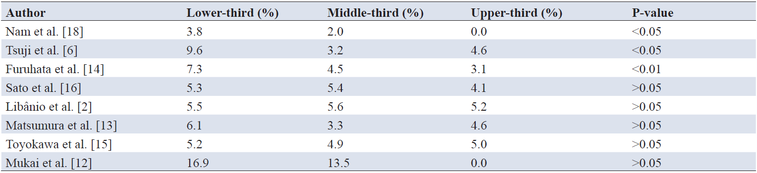
The size of the gastric lesion is a risk factor for bleeding after ESD. Theoretically, the larger the lesion, the greater the risk of bleeding after ESD [5]. In addition, there is controversy about whether tumor location affects bleeding after ESD. The ideal dissection plane is the avascular layer below the vascular network of the gastric wall and above the muscular layer [11]. Several studies reported that the bleeding rate was significantly higher for tumors located in the lower third and middle/lower third of the stomach, while other studies found that an upper location of the lesion in the stomach was a more influential risk factor [2, 6, 12-16]. Although the correlation between lesion location and bleeding after ESD is inconsistent in the literature, studies tend to support that ESD in the lower third of the stomach is associated with bleeding after ESD (Table 1) [17]. Therefore, endoscopists should more closely monitor patients with lesions located in the lower third of the stomach.

2.3 Patient-related risk factors
Studies have shown that the use of anticoagulants has a significant impact on post-ESD bleeding in the stomach. For patients receiving anticoagulant therapy, delayed bleeding after ESD remains an unpreventable adverse event [19].
The use of aspirin is also a risk factor for delayed bleeding, but there is controversy regarding the relationship between the risk of post-ESD bleeding and the aspirin treatment regimen during the procedure. A prospective study by Cho et al. on 328 patients showed that the postoperative bleeding rate in the group that continued to use aspirin was 15.6%, significantly higher than the rate of 4.2% in the group that stopped taking the drug (odds ratio [OR]=4.12, P=0.001) [20]. The authors confirmed through multivariate analysis that aspirin is an independent risk factor for post-ESD bleeding. A meta-analysis by Yang et al. covering 15 studies with 4,876 patients confirmed that the use of aspirin increases the risk of bleeding (OR=2.34, P<0.001), but subgroup analysis showed that the risk was lower with low-dose aspirin (≤100 mg/d) (OR=1.87) [21]. Similarly, a systematic review by Jaruvongvanich et al. that included a total of 2,143 patients showed through meta-analysis that the continued use of aspirin did not significantly increase the risk of bleeding (OR=1.25, P=0.20) [22]. These studies reveal the complexity of risk assessment: the absolute risk of bleeding increases by about 4–6% in patients at high risk of thrombosis who continue to take aspirin, while the risk of thrombotic events due to stopping the drug may be even higher. Therefore, when treating patients with different medication statuses, doctors need to comprehensively consider risk factors such as aspirin dosage, lesion size, and procedure time to develop an appropriate ESD plan.
Gastric ESD is increasingly being used in elderly patients, yet bleeding remains the main complication. Park et al. used the Korean National Health Insurance Service database, which included patients who underwent gastric ESD from November 2011 to December 2022 [8]. The elderly group (aged >75 years) accounted for 18.6% of all cases. The study used a mixed-effects logistic regression model to analyze and identify the risk factors for bleeding and analyzed the risk factors for bleeding in the elderly and non-elderly groups, respectively, to evaluate the differences in their patterns. In the multivariate logistic regression, significant risk factors included younger age, male sex, a higher comorbidity index, gastric cancer, multiple lesions, several comorbidities (such as renal failure), antithrombotic drug use, being underweight, smoking, and heavy drinking habits. The impact of some factors on the risk of bleeding differed significantly between age groups. In the elderly group, the risk was increased in association with a high comorbidity index (≥5). Although the incidence of bleeding was higher in the elderly group, this may be due to the higher prevalence of comorbidities and more frequent use of antithrombotic drugs in this population, rather than age itself [8]. The correlation between age and the risk of bleeding is consistent with previous studies, and doctors need to consider age when developing appropriate treatment plans for patients.
3.1 Observation without immediate intervention
For small polyps with a wound diameter of less than 2 cm after electrocoagulation or electroresection, if there is no bleeding, perforation, or other complication, generally no treatment is required. For some patients with early gastric cancer or early esophageal cancer and larger wounds with a diameter greater than 2 cm, if the muscularis propria is intact after ESD, hemostasis is effective during the procedure, the risk of perforation is low, and further intervention is difficult after the procedure, wound observation without immediate intervention can also be considered [23]. After the procedure, patients should fast, stay in bed, and receive enhanced acid suppression treatment.
Epinephrine has the effect of constricting blood vessels and achieving local hemostasis and is often added to the submucosal injection solution [24]. The injection needle consists of an outer sheath (made of plastic, polytetrafluoroethylene, or stainless steel) and an inner hollow needle. Using the handle at the end of the needle sheath, the operator uses a syringe connected to the handle to inject liquid agents into the target tissue. The injection of various solutions achieves hemostasis through mechanical tamponade and chemical mechanisms. A study showed that preoperative submucosal injection of epinephrine during EMR for colonic polyps significantly reduced the risk of bleeding after colonic polyp resection compared with no intervention, and the cost was significantly lower than that of mechanical hemostasis methods such as hemostatic clips. Hemocoagulase for injection is a hemostatic agent extracted from snake venom that can promote the formation of fibrin polymers, thereby achieving hemostasis, and is commonly used in surgical procedures.
3.2 ESD ulcer closure
Endoscopic ulcer closure and hemostasis include the use of hemostatic clips, the combination of hemostatic clips and nylon ropes for wound closure, and the use of over-the-scope clips (OTSCs). Hemostatic clips are one of the most widely used endoscopic closure devices after treatment. In areas where gastrointestinal ligation devices are difficult to use for closure, hemostatic clips are generally selected. Clinically, for small wounds, a single hemostatic clip can completely close the wound; for larger wounds, multiple hemostatic clips are needed for continuous closure similar to sutures, or a combination of hemostatic clips and nylon ropes can be used for purse-string closure. The literature reports that endoscopic purse-string suturing using hemostatic clips and nylon ropes to treat large lesions in the gastric antrum (single lesion with a diameter greater than 3 cm) is safe and feasible [25]. The OTSC system with a rake-shaped metal clip can effectively close large-diameter gastrointestinal perforations or refractory gastrointestinal fistulas. Prophylactic closure of artificial ulcer surfaces with the rake-shaped metal clip can effectively prevent postoperative perforation.
For postoperative bleeding after ESD, through-the-scope clipping (TTSC), OTSC, and suturing represent the main techniques for tissue apposition [25]. TTSC is the most commonly used hemostatic clip in clinical practice at present. It is released through the endoscopic working channel and is suitable for clamping small mucosal or submucosal bleeding points. It has the advantages of easy operation and low cost. However, its clamping force is limited, and it can usually only close superficial tissues. It has poor closure effects for deeper or larger wounds, perforations, or full-thickness defects. In contrast, OTSC is a cap-like device mounted at the front end of the endoscope. It achieves strong occlusion through a spring-loaded mechanism and can simultaneously clamp the mucosal layer, muscular layer, and even the serosal layer. It is suitable for closing full-thickness gastrointestinal wall defects (such as perforations and fistulas) or treating massive bleeding. OTSC has a larger clamping range, deeper grasping ability, and stronger closing force, but the operation is relatively complex, the cost is high, and it usually cannot be released repeatedly. TTSC is suitable for superficial hemostasis during routine ESD, while OTSC is more commonly used for the emergency treatment of complex complications, such as perforations, delayed massive bleeding, or fistulas. The two are complementary in clinical application.
3.3 ESD ulcer shielding
3.3.1 Hemostatic powders
Polyglycolic acid (PGA) is a high-molecular-weight polymer. It is a soft material with certain ductility and tensile strength and can be hydrolyzed into carbon dioxide and water. Therefore, it is safe for application in the human body. The earliest application of PGA in the treatment of wounds after digestive tract ESD was reported in 2012. Takimoto et al. used PGA combined with fibrin glue to prevent delayed perforation after ESD of duodenal tumors with a diameter of 20 mm [26]. The PGA sheet was placed over the ulcer with biopsy forceps and fixed with sprayed fibrin glue, and it was absorbed spontaneously within four to fifteen weeks. This shows that the endoscopic tissue shielding method using PGA sheets and fibrin glue has potential in preventing bleeding after ESD.
In recent years, several hemostatic powders have been developed and clinically used to treat bleeding after gastric ESD. These hemostatic powders can be used clinically (Table 2) [27]. In the study by Zhang et al., patients (n=173) who underwent ESD from January 2009 to June 2012 were included in a prospective randomized study [28]. The remaining 171 patients (excluding two patients whose procedures were aborted) were randomly divided into two groups: Group A (medical adhesive group, n=89) and Group B (control group, n=82). There were no significant differences in the average treatment time (59.4 min and 55.0 min, respectively) and the incidence of intraoperative perforation (10.1% in Group A and 9.8% in Group B) between Group A and Group B. There was a significant difference in the average length of hospital stay between Group A and Group B (8.89 days and 9.90 days), and the incidence of delayed bleeding after the procedure in Group A was significantly lower than that in Group B (0.00% and 4.88%).

One of the advantages of applying hemostatic powder is that it requires minimal technical expertise. Since the powder only needs to be sprayed onto the surface of the ulcer after ESD through a catheter, regardless of the anatomical location, hemostatic powder has certain natural advantages in difficult areas [29]. Therefore, beginners in ESD can easily apply hemostatic powder to prevent bleeding after ESD-induced iatrogenic ulcers [27]. However, there is a learning curve for those who use hemostatic powder for the first time. In addition, hemostatic powder can be quickly and easily applied to large lesions. Moreover, due to its non-contact application, hemostatic powder does not cause additional secondary tissue damage [30]. Therefore, applying hemostatic powder is an effective treatment option for protecting the ulcer surface after ESD and reducing postoperative scarring [31]. However, hemostatic powder may be affected by gravity. Most powders adhere well to the surface due to the moisture of the ulcer after ESD. After the endoscopist sprays the powder onto the target lesion, the remaining powder can accumulate in a gravity-dependent manner in the target area.
The hemostatic material TC-325 (Hemospray®, Cook Medical, Winston-Salem, NC, USA) has been reported as an endoscopic hemostatic option for gastrointestinal bleeding, including bleeding after endoscopic procedures [32, 33]. Aziz et al. studied and evaluated the efficacy of Hemospray in patients with non-variceal upper GIB [33]. The primary outcomes were clinical and technical success; the secondary outcomes were clustered rebleeding, early rebleeding, delayed rebleeding, refractory bleeding, mortality, and treatment failure. The final analysis included a total of 20 studies and 1,280 patients. Technical success of Hemospray was observed in 97% of the cases, and a significant trend of increasing technical success was observed during the publication period from 2011 to 2019. The clinical success rate of Hemospray was 91%, while the clinical success rate of other hemostatic measures was 87%. After using Hemospray, the secondary outcomes of clustered rebleeding, early rebleeding, delayed rebleeding, refractory rebleeding, mortality, and treatment failure occurred in 27%, 20%, 9%, 8%, 8%, and 31% of the cases, respectively. Hemospray is safe and effective in treating non-variceal upper GIB, is not inferior to traditional hemostatic measures, and can be used as an alternative option.
Polysaccharide hemostatic materials have been proven in multiple clinical trials to be applicable to the treatment of post-ESD bleeding. Yu et al. used a carbon dioxide insufflator (CO2 Regulation Unit, Olympus) to insufflate the hemostatic material [34]. This device uses high airflow during the catheter insertion stage to disperse moisture. After aiming the catheter at the ulcer surface, the assistant changes the mode to low airflow to evaluate the efficacy of the newly designed polyethylene oxide (PEO) adhesive in preventing delayed bleeding. The rate of delayed bleeding in the PEO group was relatively lower than that in the control group, but the difference was not significant (P=0.094). No bleeding events occurred within 48 hours after ESD in the PEO group, while the bleeding rate after 48 hours in the control group was 60.9% (14/23). Cha et al. used the hemostatic powder UI-EWD (NexpowderTM, Nextbiomedical, Incheon, South Korea) to study the endoscopic hemostasis success rate, UI-EWD-related adverse events, and rebleeding rate [35]. The incidence of endoscopic hemostasis in the UI-EWD group for lesions located at the hepatic flexure (7.3% vs. 0.0%, p=0.011) and lesions larger than 4 cm (25.5% vs. 8.0%, p=0.002) was significantly higher than that in the conventional treatment group. The cumulative rebleeding rate within 28 days was 5.5% (3/55), significantly lower than that in the conventional treatment group (17.0%, 19/112; p=0.039). For lower GIB with anatomical or technical obstacles to endoscopic access, Cha et al. believe that UI-EWD should be considered first.
3.3.2 New treatment option: self-assembling peptide TDM
The synthetic self-assembling peptide TDM is derived from non-biological materials and consists of 16 amino acid residues. It is usually in the form of an aqueous solution and can easily pass through a catheter. The self-assembling peptide can form a gel under specific physiological conditions [36]. Therefore, the self-assembling peptide TDM can be used not only in open surgery but also in flexible endoscopic treatment.
The first generation of self-assembling peptides (TDM-621, 3-D Matrix, Tokyo, Japan) is currently used as a hemostatic agent in Europe. TDM-621 has some drawbacks. Its gel formation rate is slow, and its retention on the wound is poor. Therefore, the second-generation synthetic peptide TDM-623 was developed. TDM-623 has been developed as the second generation of self-assembling peptide materials, which can accelerate gel formation and improve tissue retention. Yoshida et al. tested the feasibility of the synthetic self-assembling peptide TDM-621 for hemostasis after endoscopic treatment of gastric tumors and evaluated its hemostatic effect in 12 patients [37]. No secondary bleeding was observed in any of the 12 patients, and no adverse reactions considered to be related to TDM-621 were observed. However, this study not only has a small sample size but also lacks a direct efficacy comparison with traditional mechanical clipping or prophylactic electrocoagulation, and further verification is needed. Kubo et al. conducted an animal experiment using the self-assembling peptide TDM-623 and performed endoscopic hemostatic treatment with the self-assembling peptide on 23 bleeding lesions [36]. Seventeen of the 23 cases (74%) achieved complete hemostasis on the anterior wall of the stomach. Kubo et al. believe that the new self-assembling peptide (TDM-623) shows a strong hemostatic effect, and TDM-623 has potential practical value for upper gastrointestinal endoscopic procedures [36]. The success rate of the new-generation material in animal models (74%) is lower than that of the first-generation material in human single-arm studies (100%). This illustrates the limitations of the current study. There are huge differences in hemodynamics and microenvironment between artificial wound hemorrhage in animal models and real human postoperative ESD hemorrhage; at the same time, the remaining 26% hemostatic failure rate also indicates that the procedural protocol relying solely on self-assembling peptides to deal with high-pressure arterial hemorrhage is unreliable. On the whole, the current literature on self-assembling peptides mainly remains at the stage of small-sample case series or animal experiments. Before self-assembling peptides can be routinely applied to upper gastrointestinal endoscopic treatment, large-sample, multicenter randomized controlled trials with positive controls (such as TC-325 hemostatic powder or traditional hemostatic clips) need to be carried out to clarify their true clinical benefits and applicable boundaries.
With the development of endoscopic technology, endoscopic treatment techniques for gastric ESD provide a less invasive and effective treatment method than traditional surgery. Endoscopy is becoming the first-line method for treating precancerous lesions of gastric cancer. Although significant progress has been made in research on post-ESD bleeding in the stomach, there is still room for further research. With population aging and the increasing prevalence of chronic diseases, the use of aspirin and other anticoagulant drugs in clinical practice has increased significantly, and they have become important means for the primary and secondary prevention of cardiovascular diseases. In the face of an aging population and patients receiving anticoagulant therapy, in addition to traditional mechanical closure and thermocoagulation therapy, the development of tissue shielding technology and the emergence of new aqueous hemostatic materials provide a new solution to make up for the limitations of traditional devices in treating large-area mucosal defects.
A review of the treatment of bleeding after ESD in recent years shows that clinical practice urgently needs to shift from simple post-hemorrhage remediation to risk-based procedural management. Doctors not only need to accurately evaluate the use of antithrombotic drugs before the procedure but also need to carry out personalized prevention and treatment according to the characteristics of the lesions during the procedure. Future research should focus on developing dedicated hemostatic protocols for patients receiving anticoagulant therapy and verifying the cost-effectiveness of novel hemostatic materials through multicenter clinical trials to ultimately minimize ESD complications, improve the clinical success rate, and reduce procedural risk [38].
Author contributions
Jin Xu was responsible for the literature search, literature screening, analysis and summary of relevant studies, and drafting of the manuscript. Shiju Yan provided academic guidance, supervised the overall design and structure of the review, and critically revised the manuscript for important intellectual content. All authors read and approved the final manuscript.
Funding
This research received no external funding.
Data availability
Not applicable.
Ethics approval and consent to participate
Not applicable.
Consent for publication
Not applicable.
Competing interests
The authors declare that they have no competing interests.
Acknowledgements
Not applicable.
[1] Oda I, Suzuki H, Nonaka S, Yoshinaga S. Complications of gastric endoscopic submucosal dissection. Dig Endosc. 2013 Mar;25 Suppl 1:71-78. https://doi.org/10.1111/j.1443-1661.2012.01376.x
[2] Libânio D, Costa MN, Pimentel-Nunes P, Dinis-Ribeiro M. Risk factors for bleeding after gastric endoscopic submucosal dissection: A systematic review and meta-analysis. Gastrointest Endosc. 2016 Oct;84(4):572-586. https://doi.org/10.1016/j.gie.2016.06.033
[3] Jeon MY, Park JC, Hahn KY, Shin SK, Lee SK, Lee YC. Long-term outcomes after noncurative endoscopic resection of early gastric cancer: The optimal time for additional endoscopic treatment. Gastrointest Endosc. 2018 Apr;87(4):1003-1013.e2. https://doi.org/10.1016/j.gie.2017.10.004
[4] Zhao Y, Wang C. Long-term clinical efficacy and perioperative safety of endoscopic submucosal dissection versus endoscopic mucosal resection for early gastric cancer: An updated meta-analysis. BioMed Res Int. 2018 Jan 11;2018:3152346. https://doi.org/10.1155/2018/3152346
[5] Kataoka Y, Tsuji Y, Sakaguchi Y, Minatsuki C, Asada-Hirayama I, Niimi K, et al. Bleeding after endoscopic submucosal dissection: Risk factors and preventive methods. World J Gastroenterol. 2016 Jul 14;22(26):5927-5935. https://doi.org/10.3748/wjg.v22.i26.5927
[6] Tsuji Y, Ohata K, Ito T, Chiba H, Ohya T, Gunji T, et al. Risk factors for bleeding after endoscopic submucosal dissection for gastric lesions. World J Gastroenterol. 2010 Jun 21;16(23):2913-2917. https://doi.org/10.3748/wjg.v16.i23.2913
[7] Mujtaba S, Chawla S, Massaad JF. Diagnosis and management of non-variceal gastrointestinal hemorrhage: A review of current guidelines and future perspectives. J Clin Med. 2020 Feb 2;9(2):402. https://doi.org/10.3390/jcm9020402
[8] Park JY, Kim MS, Kim BJ, Kim JG. Analysis of risk factors for bleeding after gastric endoscopic submucosal dissection with an emphasis on age and sex differences: A nationwide population-based study. Surg Endosc. 2025 Dec;39:8418-8431. https://doi.org/10.1007/s00464-025-12201-6
[9] Koh R, Hirasawa K, Yahara S, Oka H, Sugimori K, Morimoto M, et al. Antithrombotic drugs are risk factors for delayed postoperative bleeding after endoscopic submucosal dissection for gastric neoplasms. Gastrointest Endosc. 2013 Sep;78(3):476-483. https://doi.org/10.1016/j.gie.2013.03.008
[10] Ono H, Yao K, Fujishiro M, Oda I, Uedo N, Nimura S, et al. Guidelines for endoscopic submucosal dissection and endoscopic mucosal resection for early gastric cancer (second edition). Dig Endosc. 2021 Jan;33(1):4-20. https://doi.org/10.1111/den.13883
[11] Toyonaga T, Nishino E, Man-I M, East JE, Azuma T. Principles of quality controlled endoscopic submucosal dissection with appropriate dissection level and high quality resected specimen. Clin Endosc. 2012 Nov;45(4):362-374. https://doi.org/10.5946/ce.2012.45.4.362
[12] Mukai S, Cho S, Kotachi T, Shimizu A, Matuura G, Nonaka M, et al. Analysis of delayed bleeding after endoscopic submucosal dissection for gastric epithelial neoplasms. Gastroenterol Res Pract. 2012;2012:875323. https://doi.org/10.1155/2012/875323
[13] Matsumura T, Arai M, Maruoka D, Okimoto K, Minemura S, Ishigami H, et al. Risk factors for early and delayed post-operative bleeding after endoscopic submucosal dissection of gastric neoplasms, including patients with continued use of antithrombotic agents. BMC Gastroenterol. 2014 Oct 3;14:172. https://doi.org/10.1186/1471-230X-14-172
[14] Furuhata T, Kaise M, Hoteya S, Iizuka T, Yamada A, Nomura K, et al. Postoperative bleeding after gastric endoscopic submucosal dissection in patients receiving antithrombotic therapy. Gastric Cancer. 2017 Jan;20(1):207-214. https://doi.org/10.1007/s10120-015-0588-7
[15] Toyokawa T, Inaba T, Omote S, Okamoto A, Miyasaka R, Watanabe K, et al. Risk factors for perforation and delayed bleeding associated with endoscopic submucosal dissection for early gastric neoplasms: Analysis of 1123 lesions. J Gastroenterol Hepatol. 2012 May;27(5):907-912. https://doi.org/10.1111/j.1440-1746.2011.07039.x
[16] Sato C, Hirasawa K, Koh R, Ikeda R, Fukuchi T, Kobayashi R, et al. Postoperative bleeding in patients on antithrombotic therapy after gastric endoscopic submucosal dissection. World J Gastroenterol. 2017 Aug 14;23(30):5557-5566. https://doi.org/10.3748/wjg.v23.i30.5557
[17] Liu L, Liu H, Feng Z. A narrative review of postoperative bleeding in patients with gastric cancer treated with endoscopic submucosal dissection. J Gastrointest Oncol. 2022 Feb;13(1):413-425. https://doi.org/10.21037/jgo-21-466
[18] Nam HS, Choi CW, Kim SJ, Kim HW, Kang DH, Park SB, et al. Risk factors for delayed bleeding by onset time after endoscopic submucosal dissection for gastric neoplasm. Sci Rep. 2019 Feb 25;9(1):2674. https://doi.org/10.1038/s41598-019-39381-1
[19] Tomida H, Yoshio T, Igarashi K, Morita Y, Oda I, Inoue T, et al. Influence of anticoagulants on the risk of delayed bleeding after gastric endoscopic submucosal dissection: A multicenter retrospective study. Gastric Cancer. 2021 Jan;24(1):179-189. https://doi.org/10.1007/s10120-020-01105-0
[20] Cho SJ, Choi IJ, Kim CG, Lee JY, Nam BH, Kwak MH, et al. Aspirin use and bleeding risk after endoscopic submucosal dissection in patients with gastric neoplasms. Endoscopy. 2012 Feb;44(2):114-121. https://doi.org/10.1055/s-0031-1291459
[21] Yang C, Qiu Y, Li X, Shi R. Bleeding after endoscopic submucosal dissection of gastric lesions. J Dig Dis. 2020 Mar;21(3):139-146. https://doi.org/10.1111/1751-2980.12850
[22] Jaruvongvanich V, Sempokuya T, Wijarnpreecha K, Ungprasert P. Continued versus interrupted aspirin use and bleeding risk after endoscopic submucosal dissection of gastric neoplasms: A meta-analysis. Ann Gastroenterol. 2018 May-Jun;31(3):344-349. https://doi.org/10.20524/aog.2018.0251
[23] Paspatis GA, Arvanitakis M, Dumonceau JM, Barthet M, Saunders B, Turino SY, et al. Diagnosis and management of iatrogenic endoscopic perforations: ESGE position statement. Endoscopy. 2020 Sep;52(9):792-810. https://doi.org/10.1055/a-1222-3191
[24] Conway JD, Adler DG, Diehl DL, Farraye FA, Kantsevoy SV, Kaul V, et al. Endoscopic hemostatic devices. Gastrointest Endosc. 2009 May;69(6):987-996. https://doi.org/10.1016/j.gie.2008.12.251
[25] Papaefthymiou A, Norton B, Telese A, Murray C, Murino A, Johnson G, et al. Endoscopic suturing and clipping devices for defects in the GI tract. Best Pract Res Clin Gastroenterol. 2024 Jun;70:101915. https://doi.org/10.1016/j.bpg.2024.101915
[26] Takimoto K, Toyonaga T, Matsuyama K. Endoscopic tissue shielding to prevent delayed perforation associated with endoscopic submucosal dissection for duodenal neoplasms. Endoscopy. 2012;44 Suppl 2 UCTN:E414-E415. https://doi.org/10.1055/s-0032-1325739
[27] Jung DH, Park JC. Strategies that reduce post-endoscopic submucosal dissection bleeding. Korean J Helicobacter Up Gastrointest Res. 2021;21(3):194-202. https://doi.org/10.7704/kjhugr.2021.0028
[28] Zhang Y, Chen Y, Qu C, Zhou M, Ni Q, Xu L. Effects of medical adhesives in prevention of complications after endoscopic submucosal dissection. World J Gastroenterol. 2013 May;19(17):2704-2708. https://doi.org/10.3748/wjg.v19.i17.2704
[29] Baracat FI, de Moura DTH, Brunaldi VO, Tranquillini CV, Baracat R, Sakai P, et al. Randomized controlled trial of hemostatic powder versus endoscopic clipping for non-variceal upper gastrointestinal bleeding. Surg Endosc. 2020 Jan;34(1):317-324. https://doi.org/10.1007/s00464-019-06769-z
[30] Karna R, Deliwala S, Ramgopal B, Mohan BP, Kassab L, Becq A, et al. Efficacy of topical hemostatic agents in malignancy-related GI bleeding: A systematic review and meta-analysis. Gastrointest Endosc. 2023 Feb;97(2):202-208.e8. https://doi.org/10.1016/j.gie.2022.07.033
[31] Beye B, Barret M, Alatawi A, Beuvon F, Nicco C, Pratico CA, et al. Topical hemostatic powder promotes reepithelialization and reduces scar formation after extensive esophageal mucosal resection. Dis Esophagus. 2016 Aug;29(6):520-527. https://doi.org/10.1111/dote.12378
[32] Pittayanon R, Khongka W, Linlawan S, Thungsuk R, Aumkaew S, Teeratorn N, et al. Hemostatic powder vs standard endoscopic treatment for gastrointestinal tumor bleeding: A multicenter randomized trial. Gastroenterology. 2023 Sep;165(3):762-772.e2. https://doi.org/10.1053/j.gastro.2023.05.042
[33] Aziz M, Weissman S, Mehta TI, Hassan S, Khan Z, Fatima R, et al. Efficacy of Hemospray in non-variceal upper gastrointestinal bleeding: A systematic review with meta-analysis. Ann Gastroenterol. 2020 Mar-Apr;33(2):145-154. https://doi.org/10.20524/aog.2020.0448
[34] Yu Y, Hu T, Kuai X, Liu X, Li R, Zhou C. Propensity score matching analysis to evaluate efficacy of polyethylene oxide adhesive on preventing delayed bleeding after gastric endoscopic submucosal dissection. Sci Rep. 2022 Mar 16;12(1):4538. https://doi.org/10.1038/s41598-022-08499-0
[35] Cha B, Lee D, Shin J, Park JS, Kwon GS, Kim H. Hemostatic efficacy and safety of UI-EWD in lower gastrointestinal bleeding. BMC Gastroenterol. 2022 Apr 7;22(1):170. https://doi.org/10.1186/s12876-022-02247-4
[36] Kubo Y, Kobayashi S, Yamamoto K, Nakagawa Y, Yamashita K, Saito T, et al. Endoscopic application of novel hemostatic material for upper gastrointestinal bleeding. DEN Open. 2021 Aug 22;2(1):e25. https://doi.org/10.1002/deo2.25
[37] Yoshida M, Goto N, Kawaguchi M, Koyama H, Kuroda J, Kitahora T, et al. Initial clinical trial of a novel hemostat, TDM-621, in endoscopic treatments of gastric tumors. J Gastroenterol Hepatol. 2014 Dec;29 Suppl 4:77-79. https://doi.org/10.1111/jgh.12798
[38] Cereatti F, Grassia R, Drago A, Conti CB, Donatelli G. Endoscopic management of gastrointestinal leaks and fistulae. World J Gastroenterol. 2020 Aug 7;26(29):4198-4217. https://doi.org/10.3748/wjg.v26.i29.4198
ISSN: 2957-5478
Volume 4, Issue 2
June 2026
Pages: 77-147


 Log In

 On This Page
 CITE
CITE